Surgeons/Restorative dentists:
Dr. Nazario Russo – Cagliari, Italy
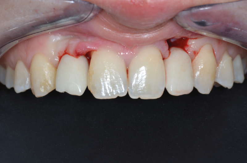
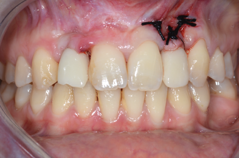
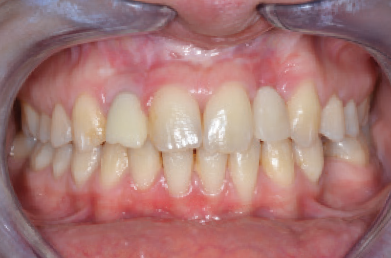
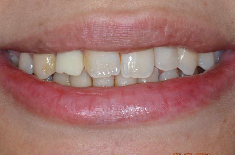
This case shows the replacement of congenitally missing maxillary lateral incisors with Narrow implants after orthodontic treatment for a 21-year-old female. Initially the anatomical conditions were ideal for inserting an XCN® Narrow 2.9 x 12 mm implant only in position #12. After 4 months of submerged healing it was decided to intervene also in position #22 and proceed with the insertion of an XCN® Narrow 2.9 x 10 mm implant opting for immediate loading with a screw-retained crown on ExaConnect. At the same time the implant in position #12 was uncovered and restored as well with a temporary screw-retained crown on ExaConnect. 2-months clinical follow-up shows an excellent restoration of aesthetics and healthy peri-implant tissues.
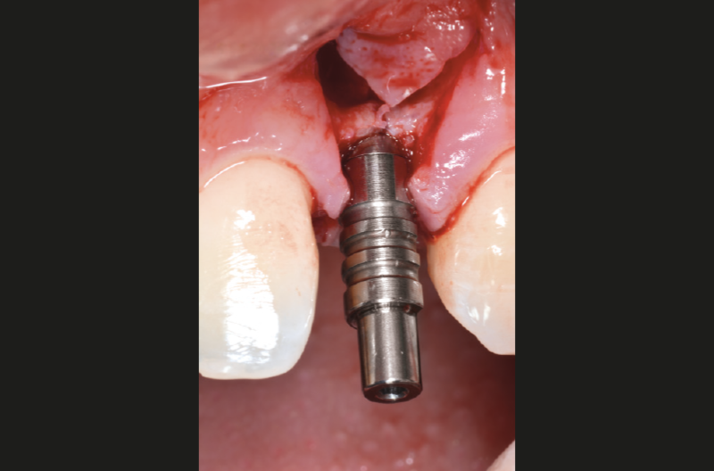
ExaConnect is a connector fixed through the Morse taper to the implant which guarantees a strong, stable and hermetically sealed connection at bone level. ExaConnect shifts the prosthetic connection from bone level to tissue level; once connected to the implant, it is no longer necessary to remove it. In position #12 ExaConnect was seated at uncovering in the implant and in position #22 at time of implant placement: in this way provisional crown fabrication, impression taking, fabrication and seating of definitive crown are done tissue level on ExaConnect, without disturbing the mucosal seal.

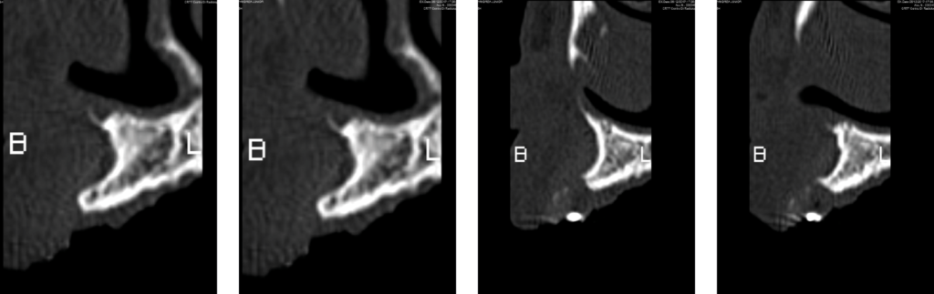
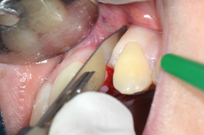
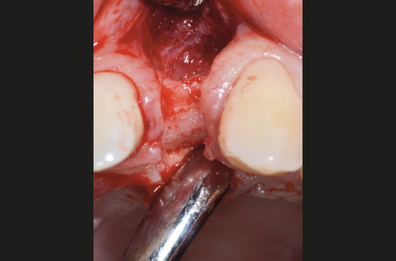
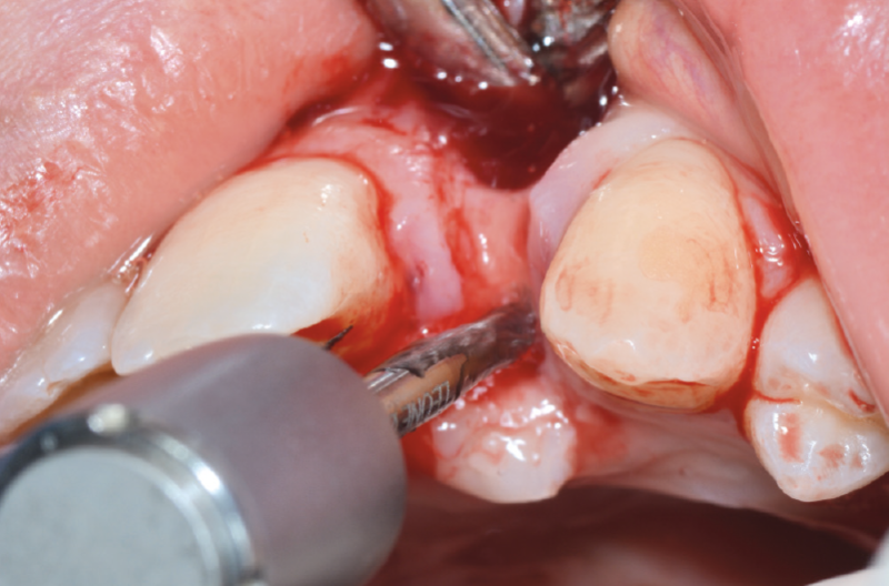
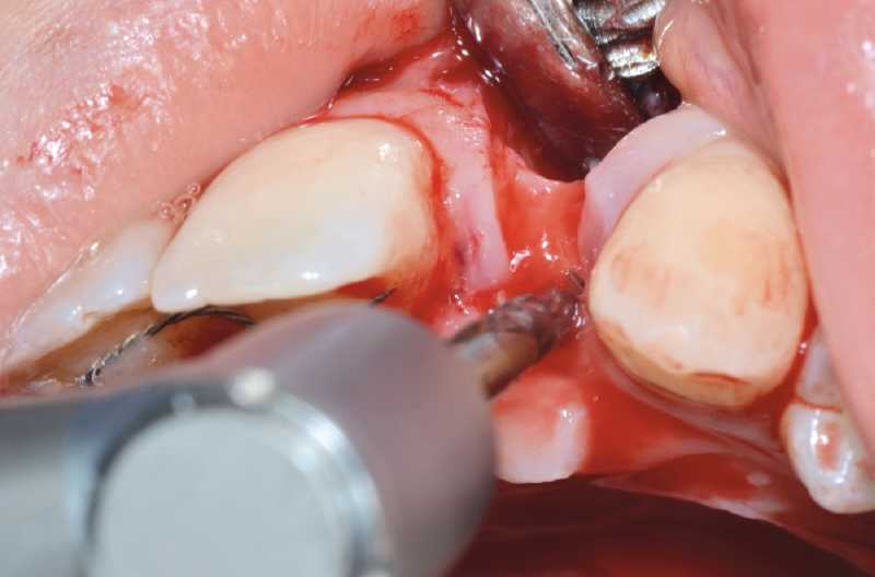
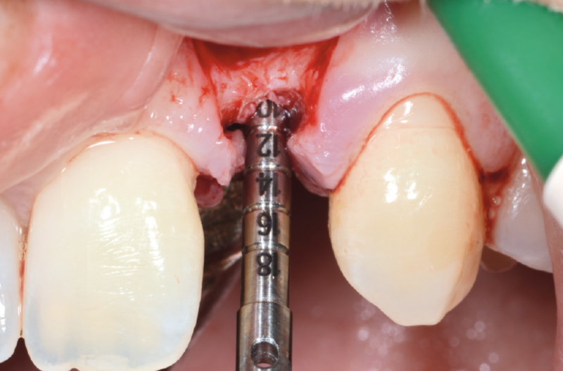
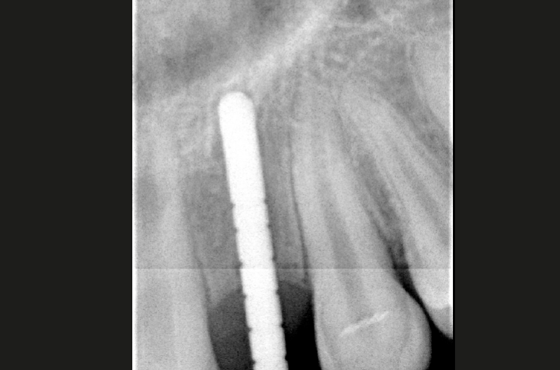
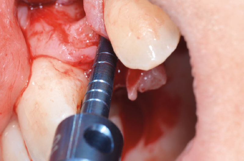
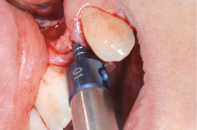
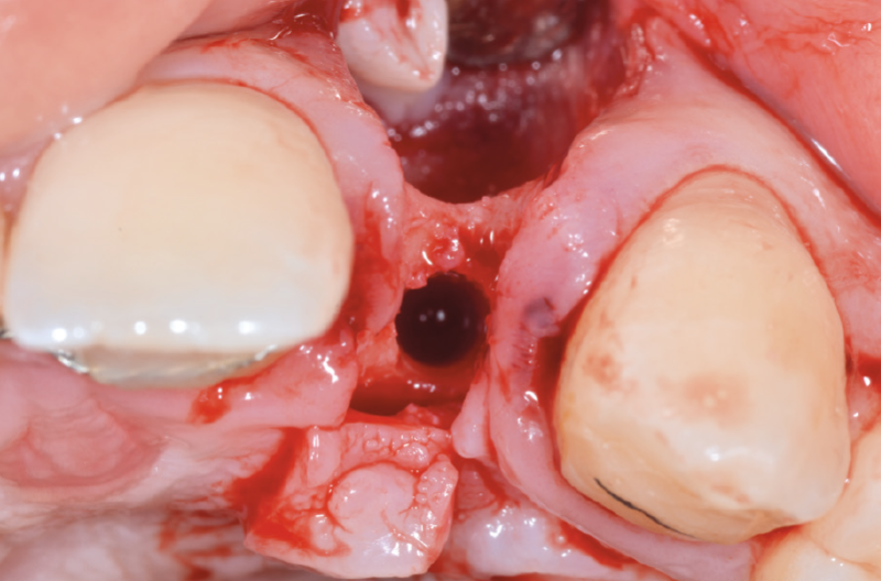
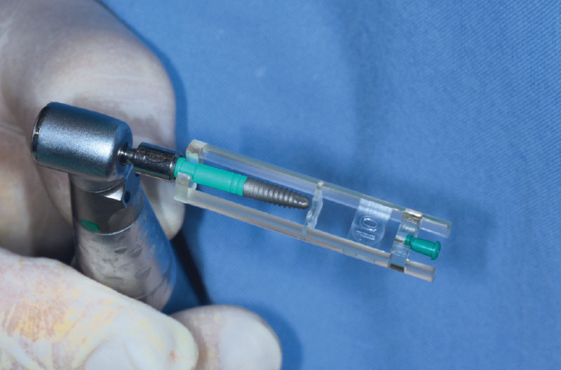
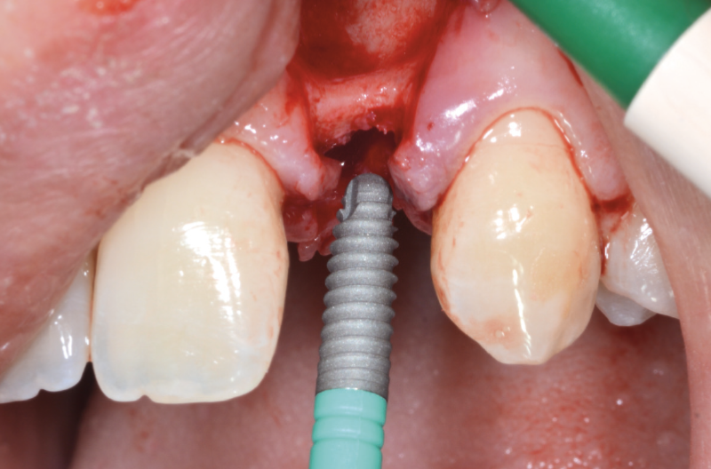
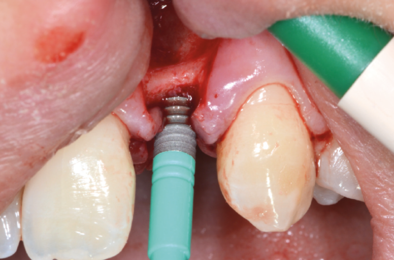
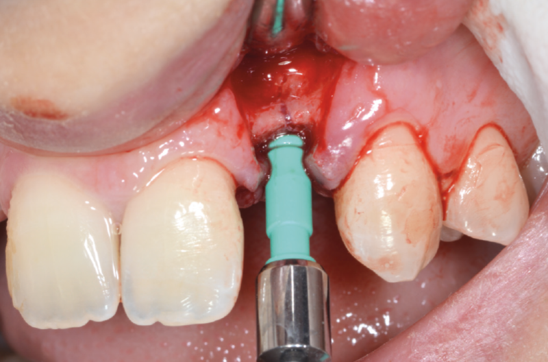
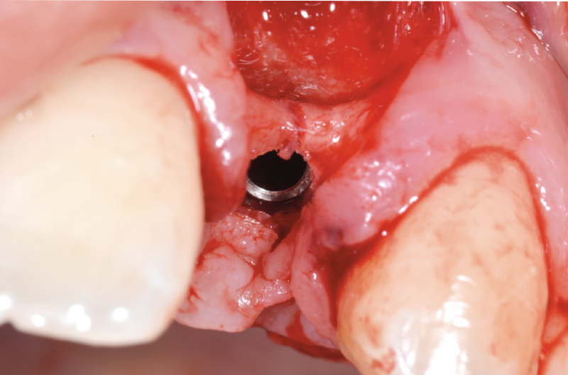
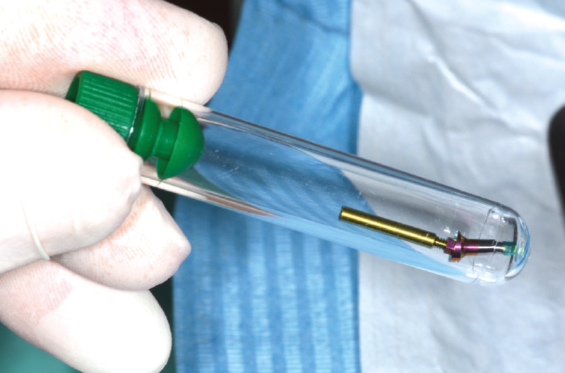
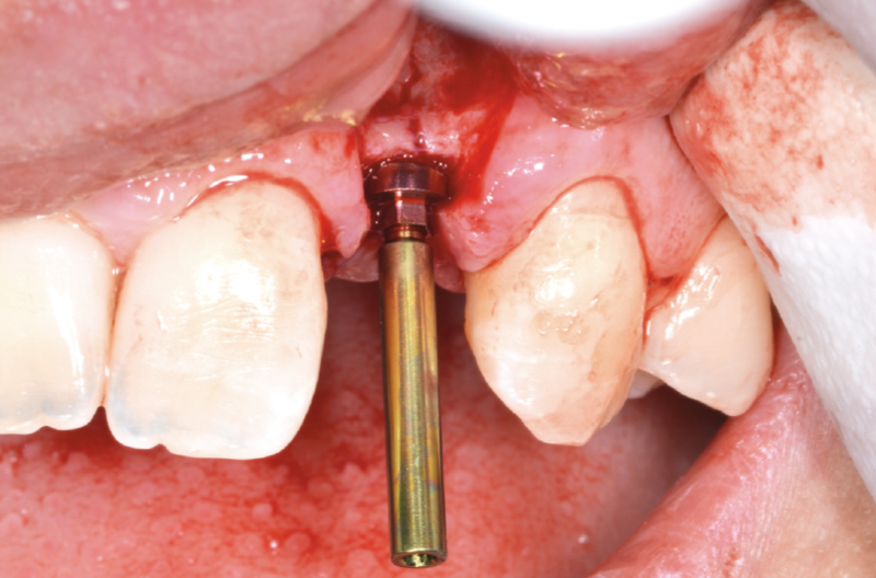
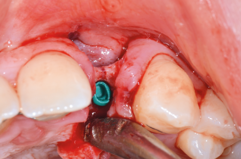
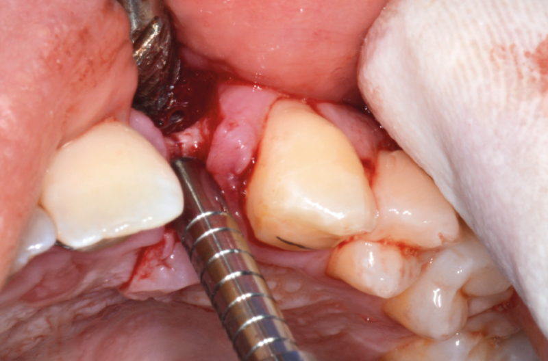
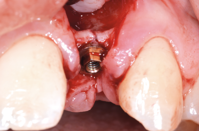
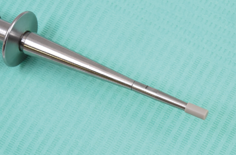
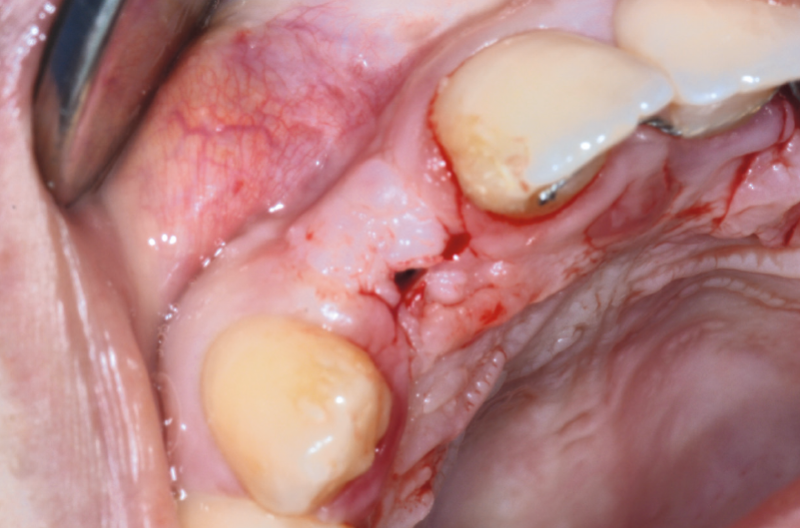
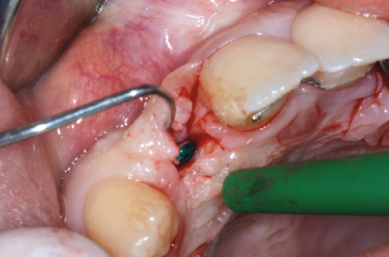
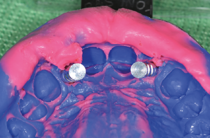
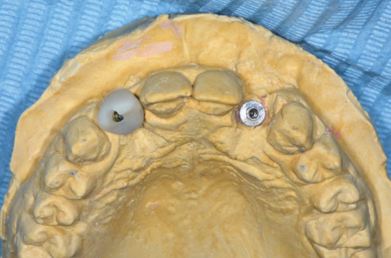
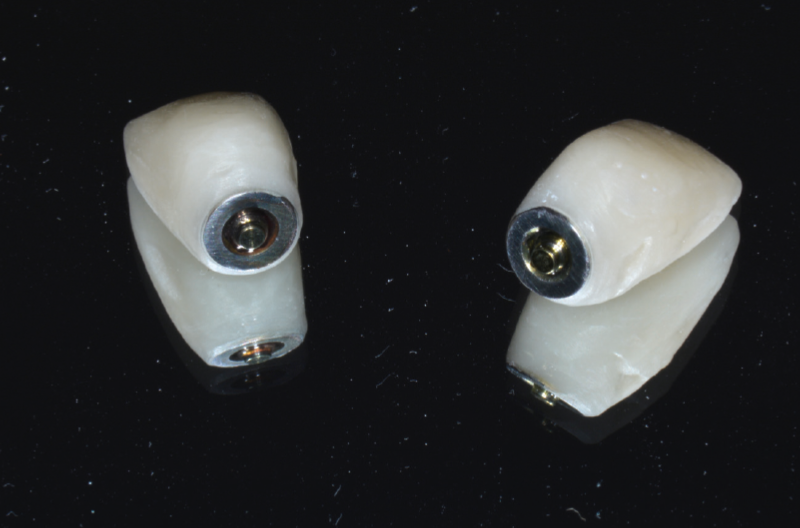
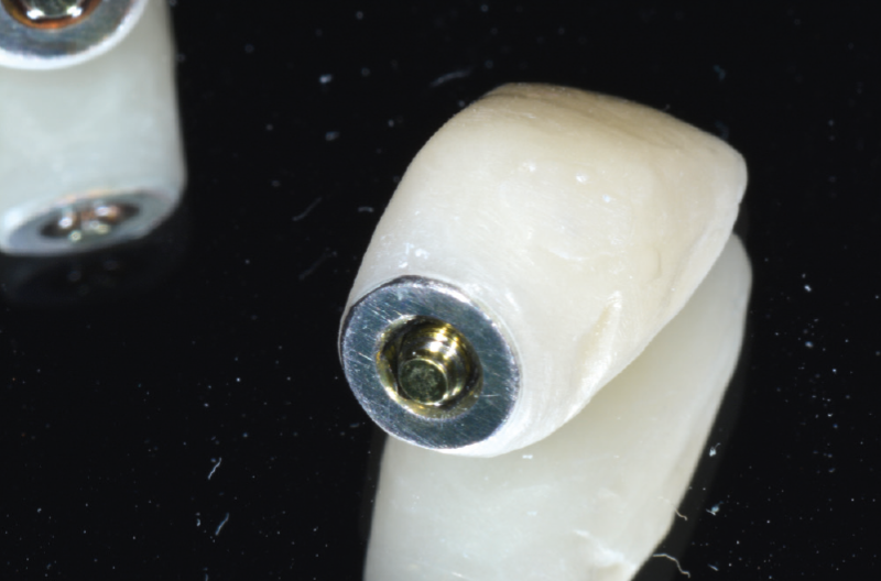
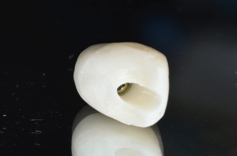
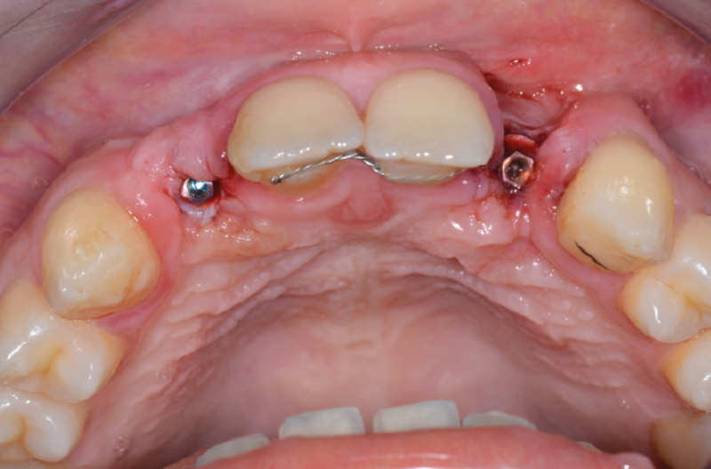
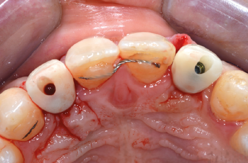
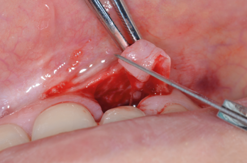
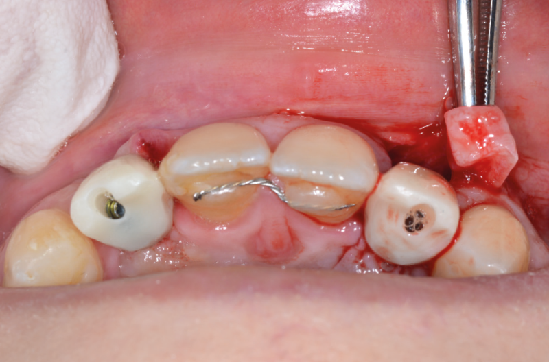
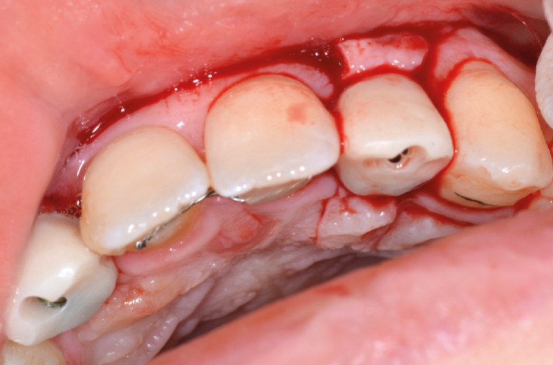
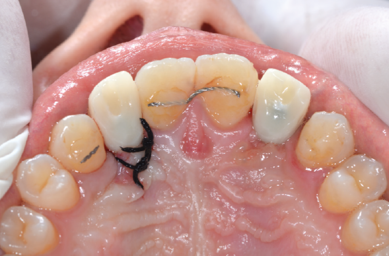
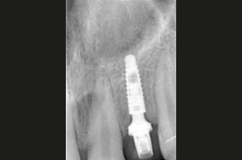
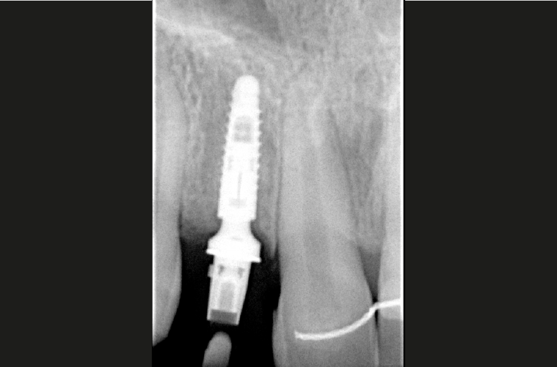
CBCT cross-section images of position #22 Rectangular full-thickness tissue flap to expose the area leaving papilla intact Rectangular full-thickness tissue flap to expose the area leaving papilla intact Creation of access hole to the bone ridge with lance drill in a more palatal position due to the presence of a large buccal concavity Use of 2.2 mm pilot drill for 11 mm Depth gauge seated in the pilot hole to take an X-ray X-ray to confirm proper distance to adjacent teeth Creation of the implant bed with a conical osteotome (2.8 mm with 2.2 tip) Creation of the implant bed with a conical osteotome (2.8 mm with 2.2 tip) View of created osteotomy Holder with a 2.9 x 10 implant coupled with its carrier Implant placement with contra-angle handpiece Implant placement with contra-angle handpiece Implant placement with contra-angle handpiece Implant placed about 1 mm subcrestally 15° angled GH 1.5 mm ExaConnect (green colour code, 2.2 connection) mounted on multifunctional screw; before activating the ExaConnect into the implant the 360° hexagon was removed View of ExaConnect in place: crestal bone margins prevent the ExaConnect from being properly seated in the implant as its prosthetic platform has a diameter of 4.1 mm and the implant is placed subcrestally View of 2.9 mm implant temporarily closed with its cover cap Use of 4.1 mm osteotome to widen the crestal bone; in this particular case the surgeon decided to use the osteotome instead of the Bone Profiler to remove bone coronally to the implant View of correctly seated ExaConnect Abutment seater with PEEK tip used to tap the ExaConnect into the well of the implant Uncovering of implant in position #12 Uncovering of implant in position #12 Pick-up transfer screwed on ExaConnect in position #22. Impression taking on both ExaConnects with open tray technique ExaConnect analogs seated on Pick-up transfers ExaConnect analogs embedded within the dental cast; #12 try-in of temporary crown on the dental cast Finished temporary screw-retained crowns Finished temporary screw-retained crowns View of the screw channel View of clinical situation a few hours after surgery Temporary crowns screwed on ExaConnects Occlusal view of temporary crowns Roll flap technique for buccal soft tissue improvement in position #22 Roll flap technique for buccal soft tissue improvement in position #22 Roll flap technique for buccal soft tissue improvement in position #22 Roll flap technique for buccal soft tissue improvement in position #22 Occlusal view of temporary crowns after closing the screw access holes with composite resin Control X-ray position #22 confirms correct placement of the implant, ExaConnect and the screw-retained crown Control X-ray position #12 confirms correct placement of the implant, ExaConnect and the screw-retained crown Clinical situation after 2 months: excellent restoration of aesthetics and healthy peri-implant tissues Clinical situation after 2 months: excellent restoration of aesthetics and healthy peri-implant tissues
Laboratory:
Gerardo Senatore – Cava De’ Tirreni (SA)