Surgeon/Restorative dentist:
Dr. Nazario Russo – Cagliari, Italy
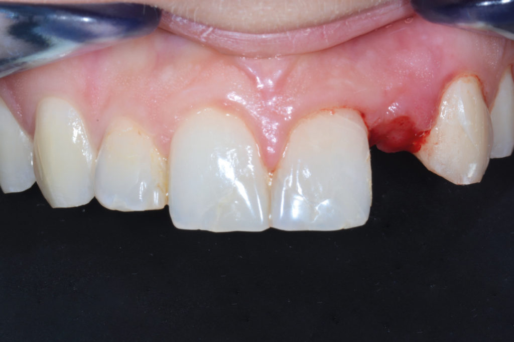
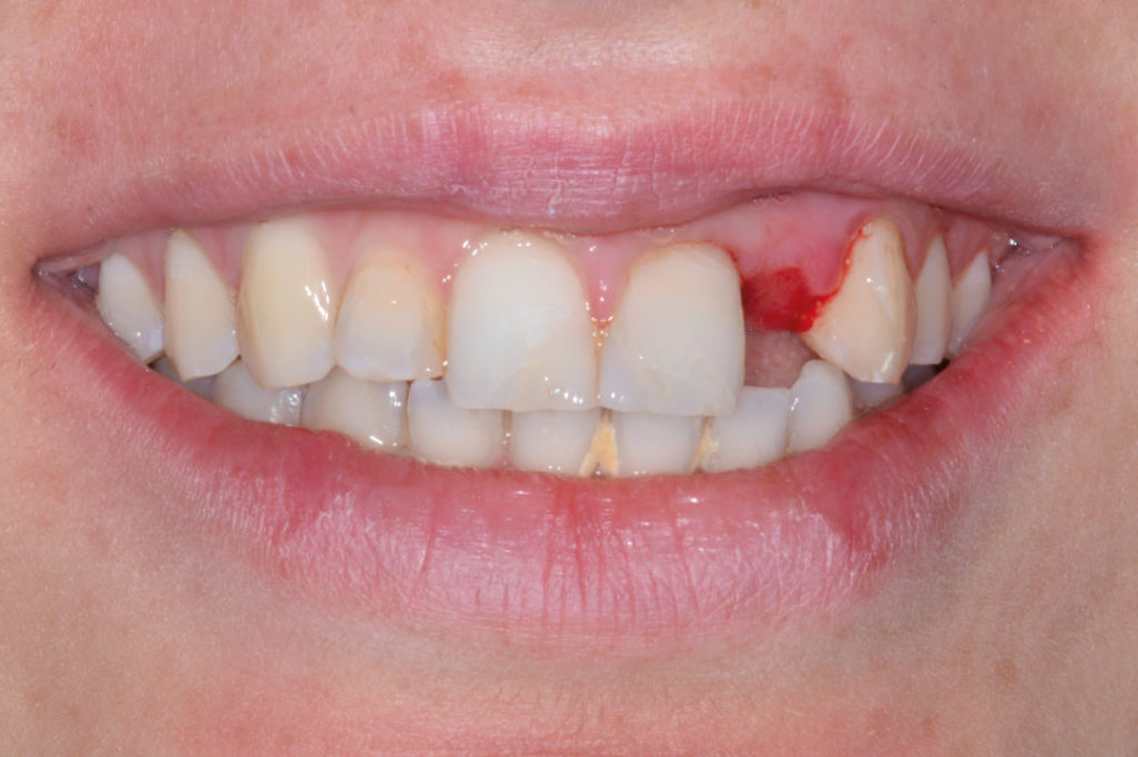
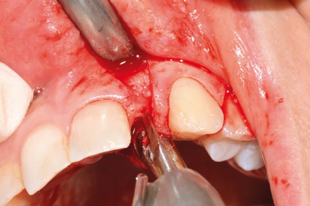
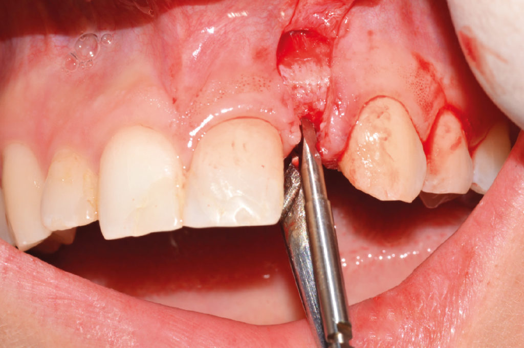
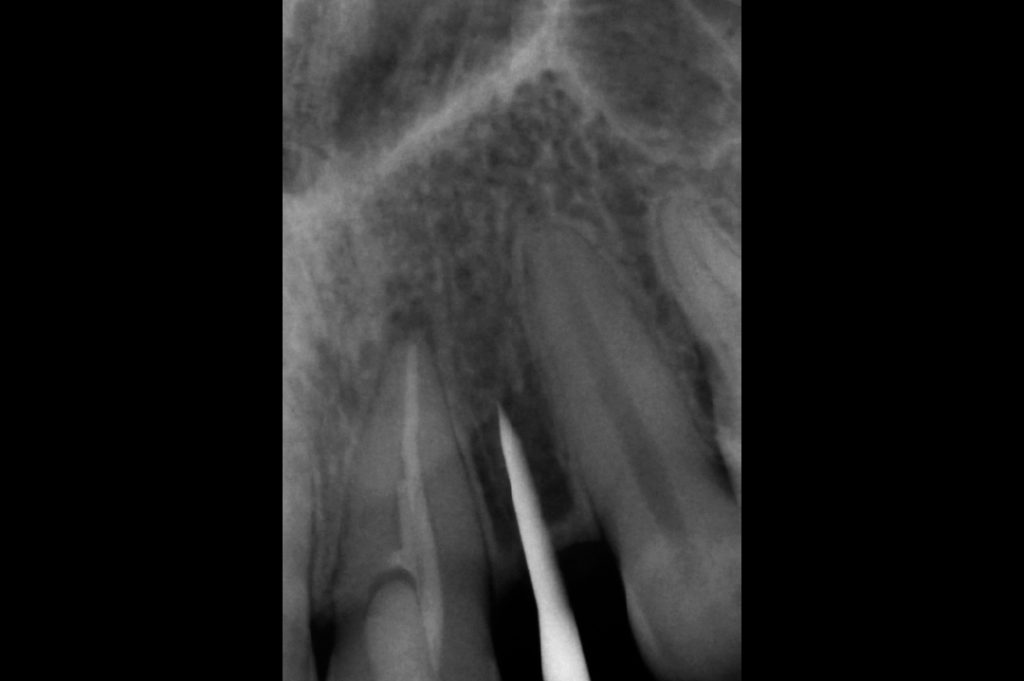
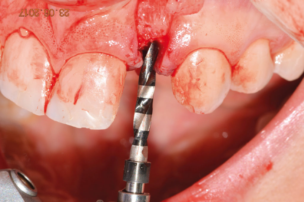
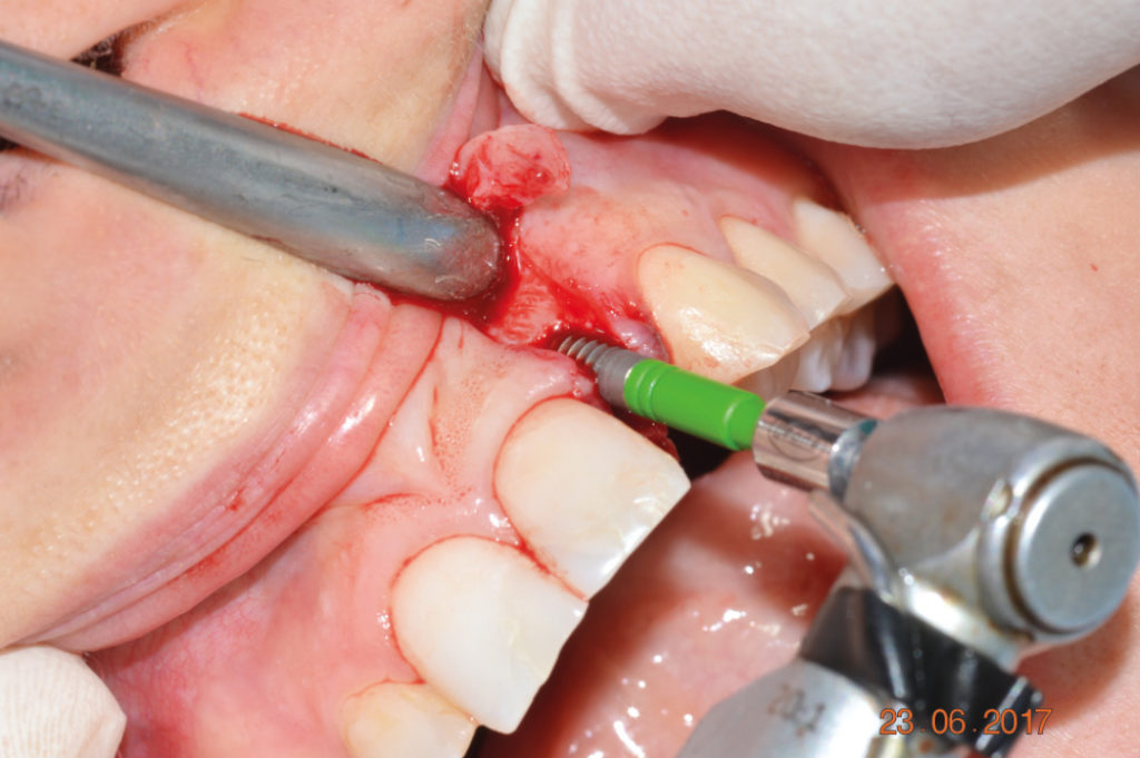
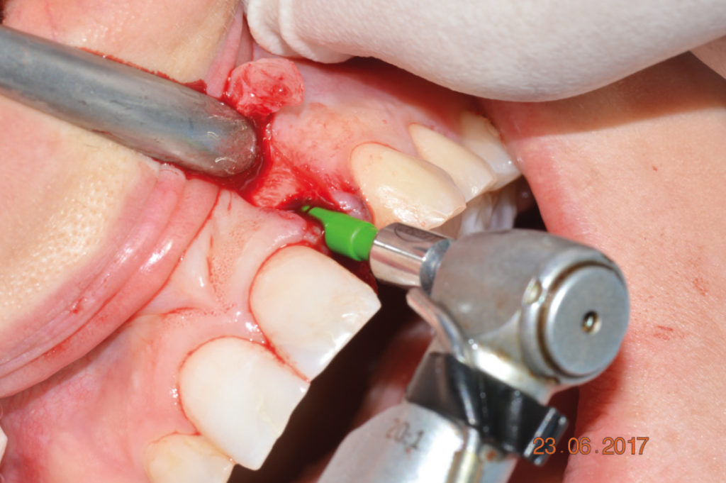
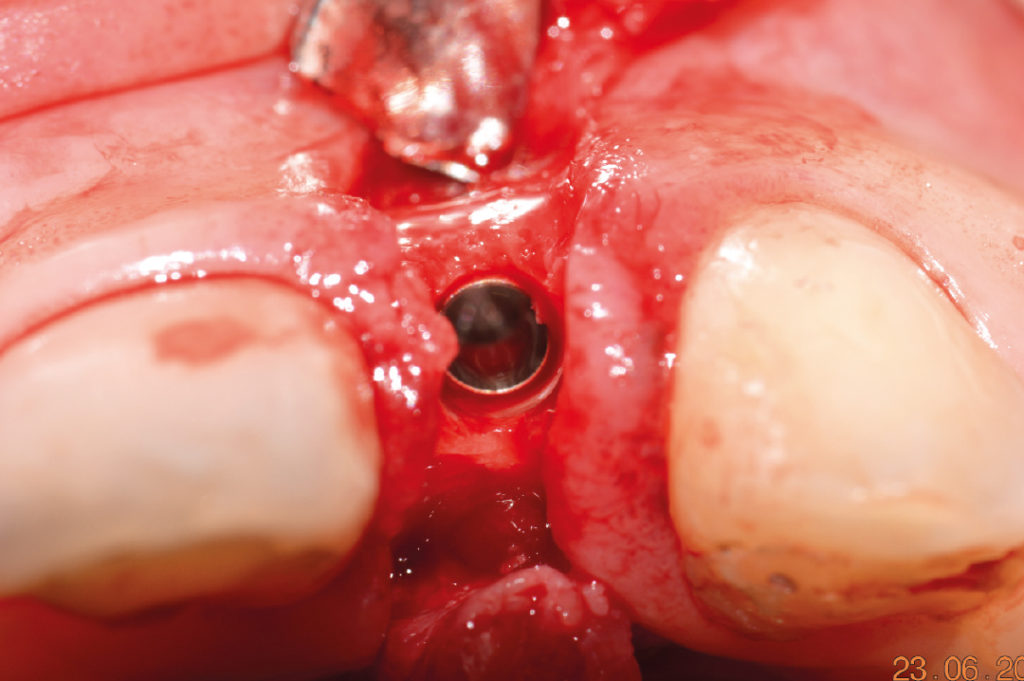
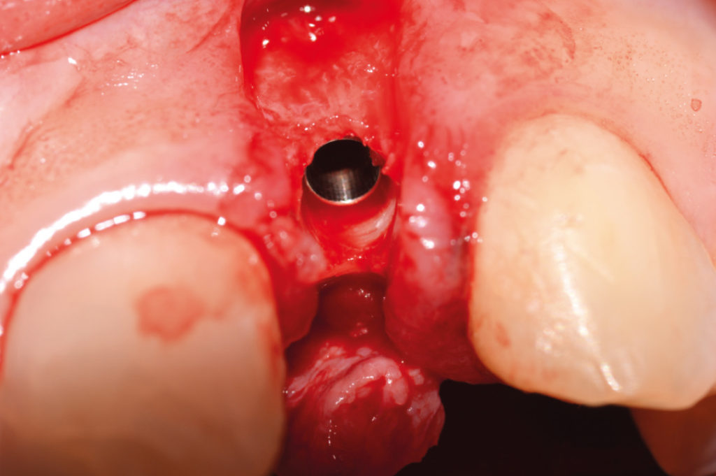
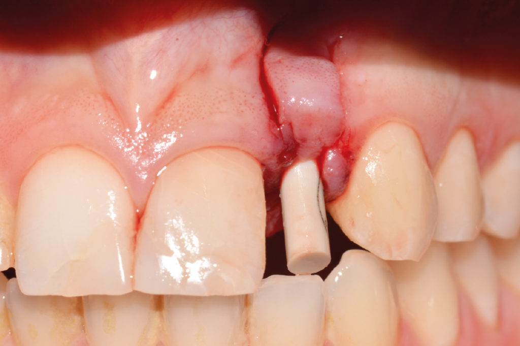
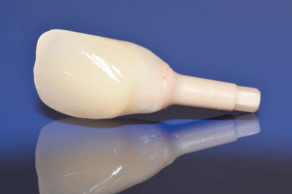
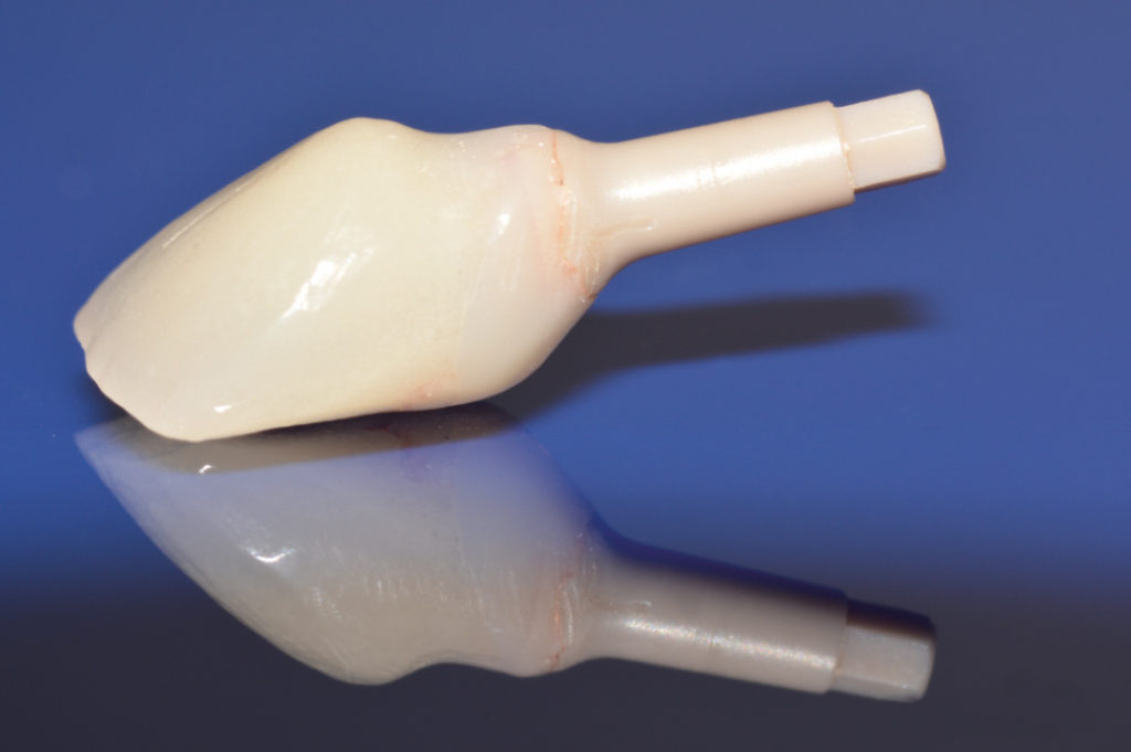
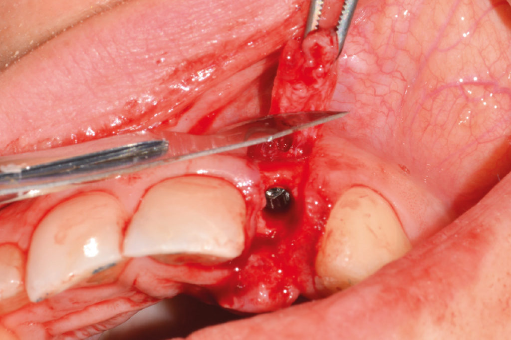
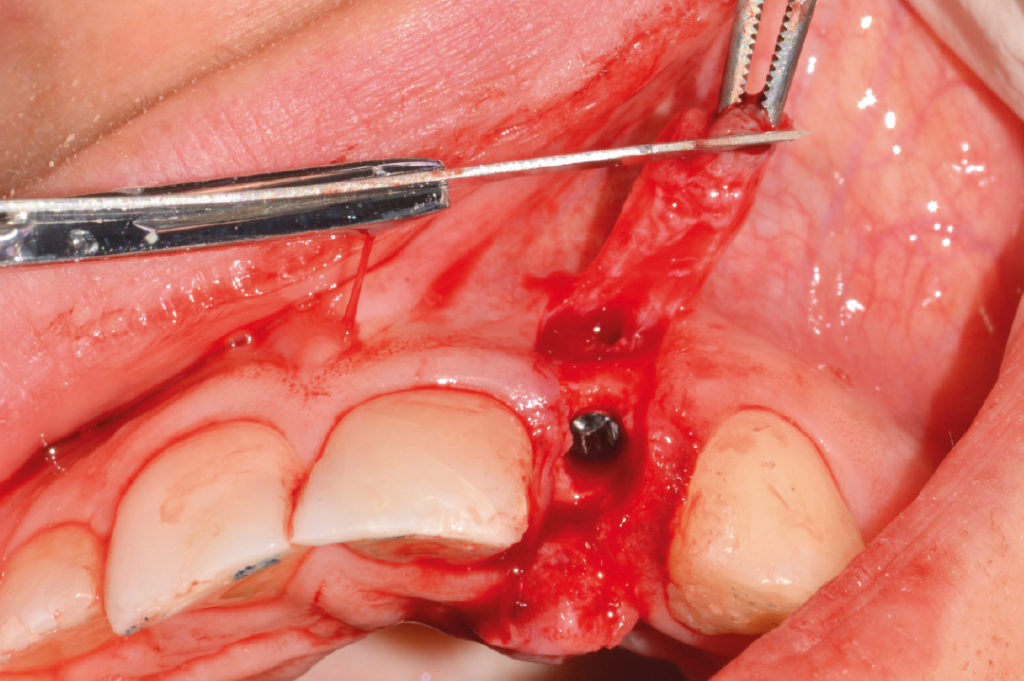
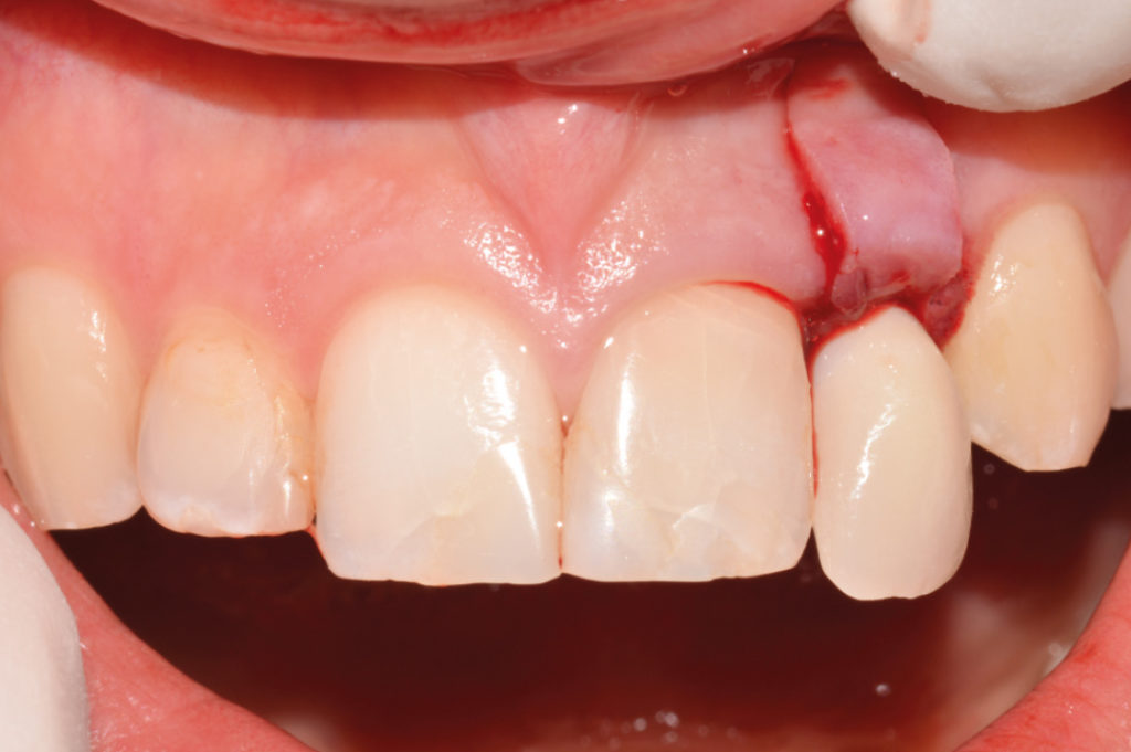
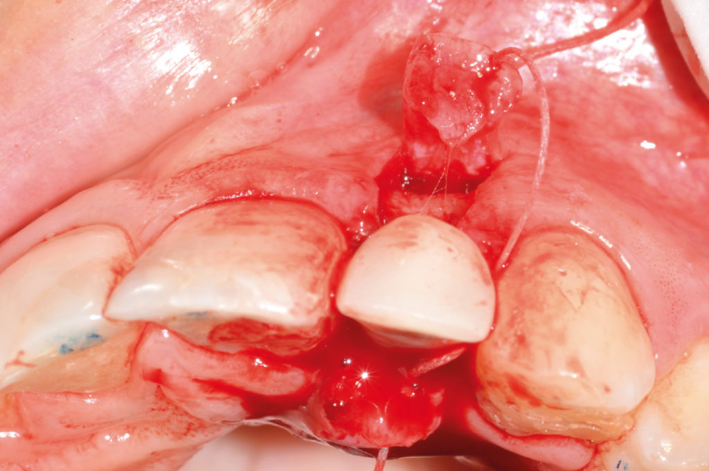
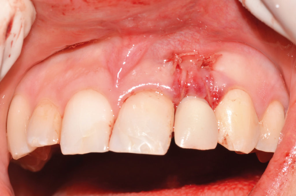
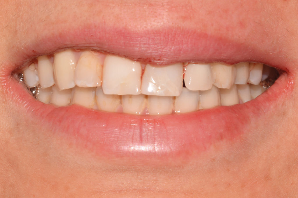
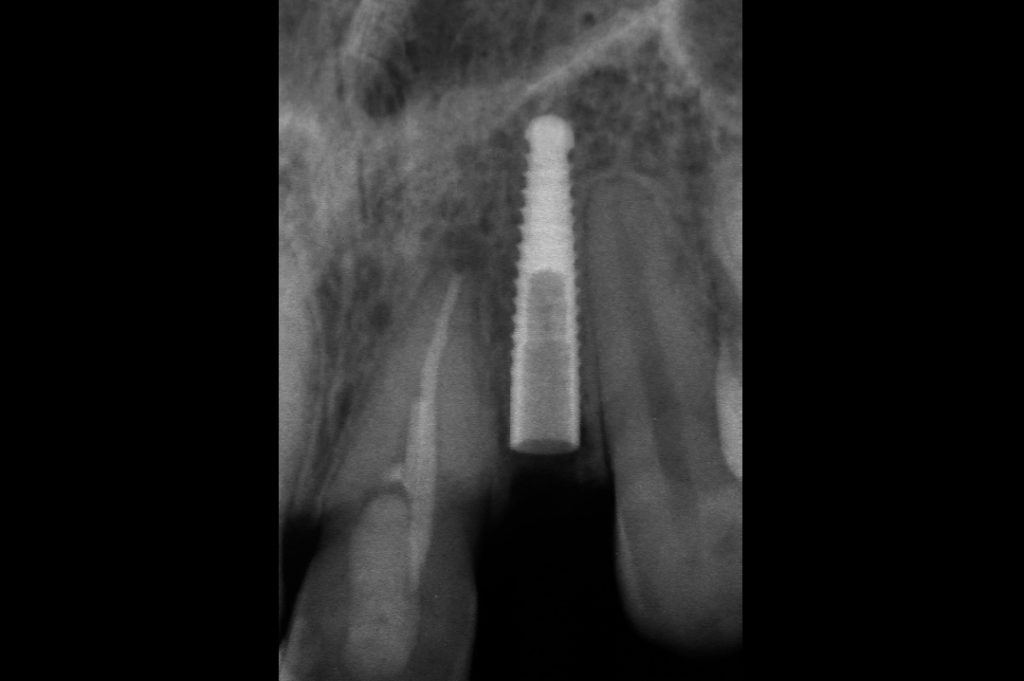
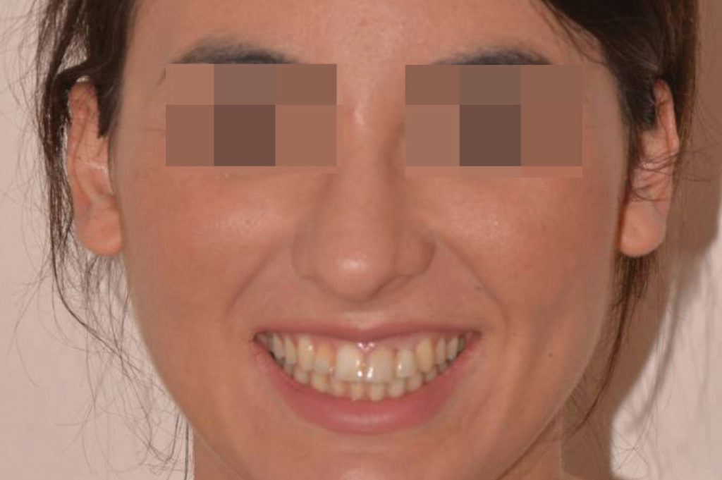
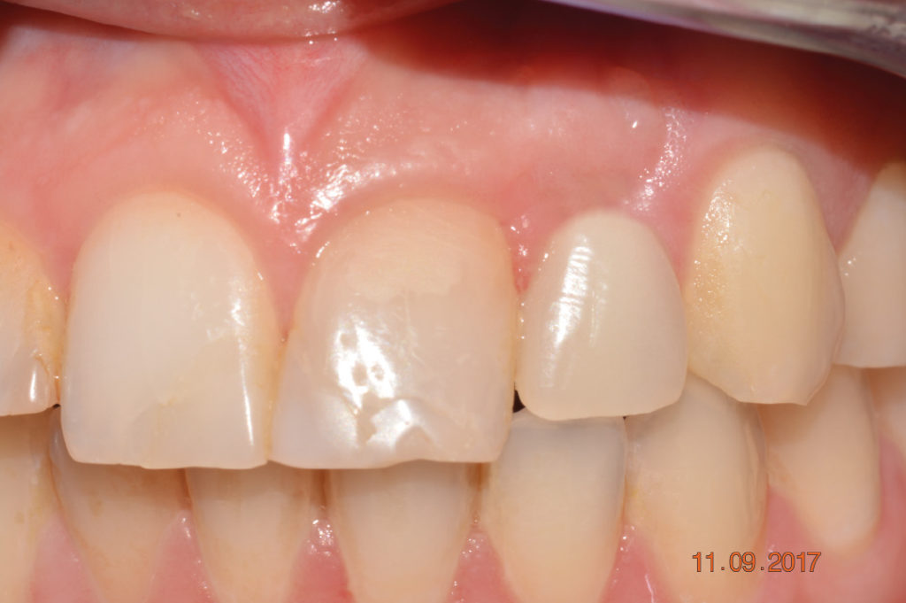
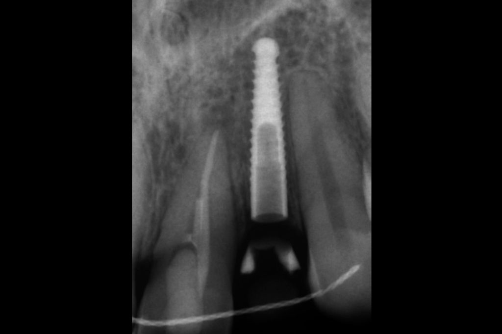
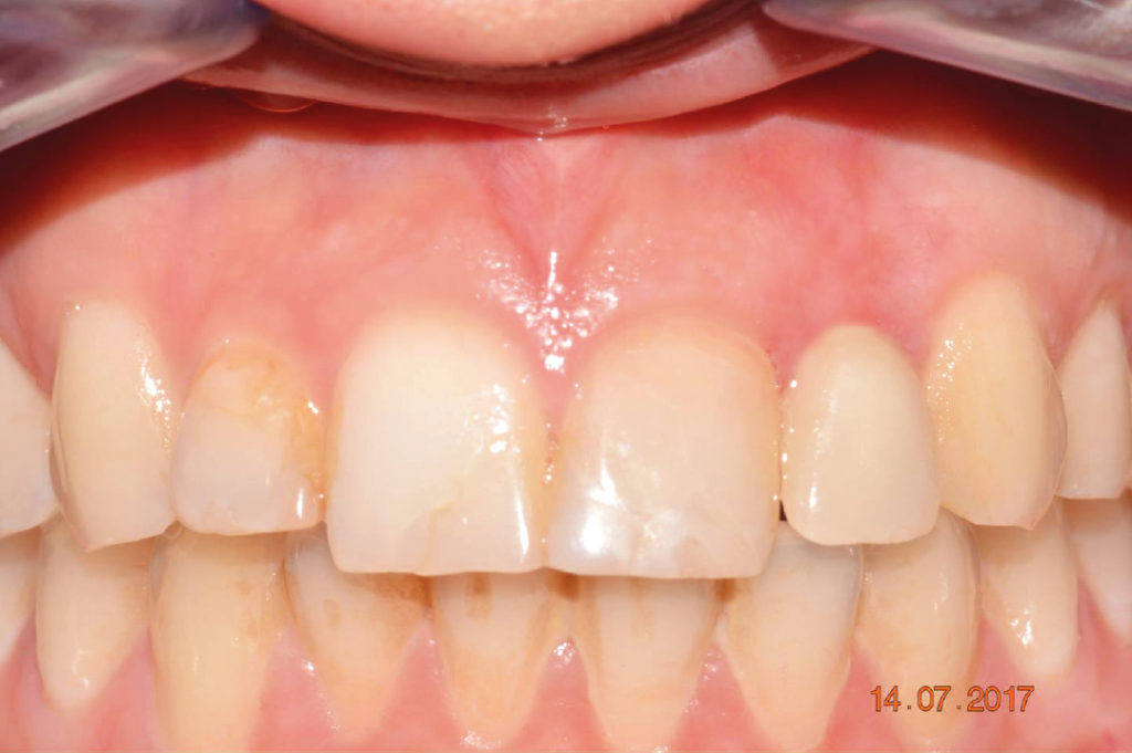
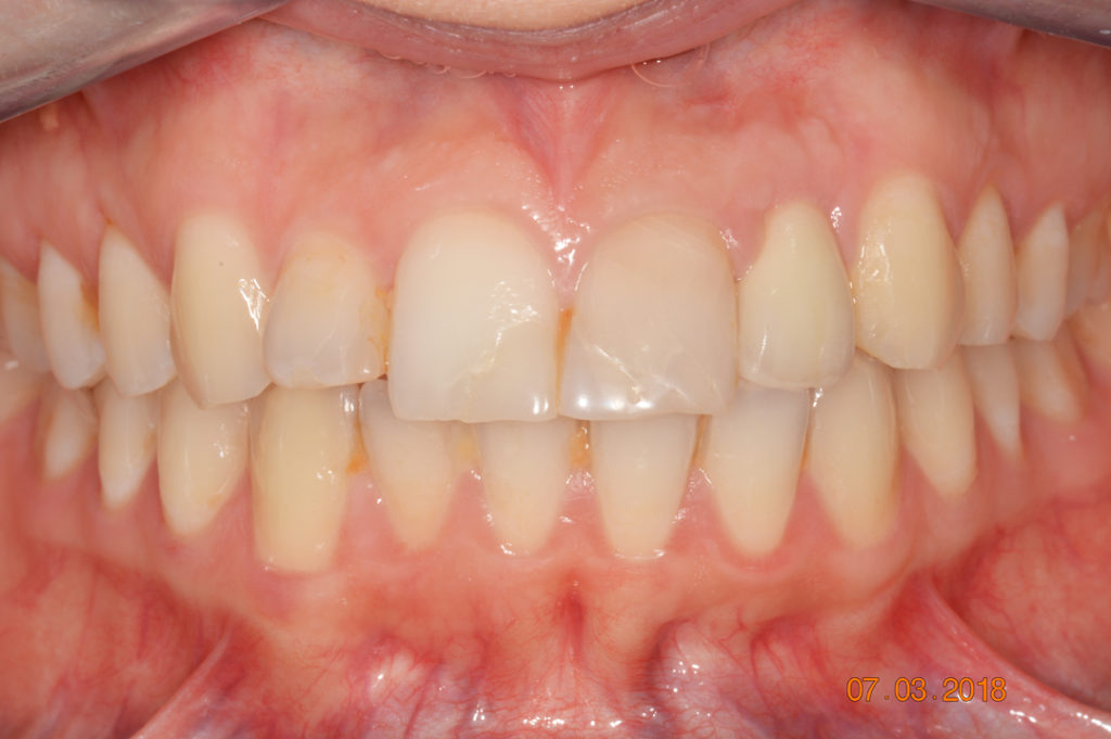
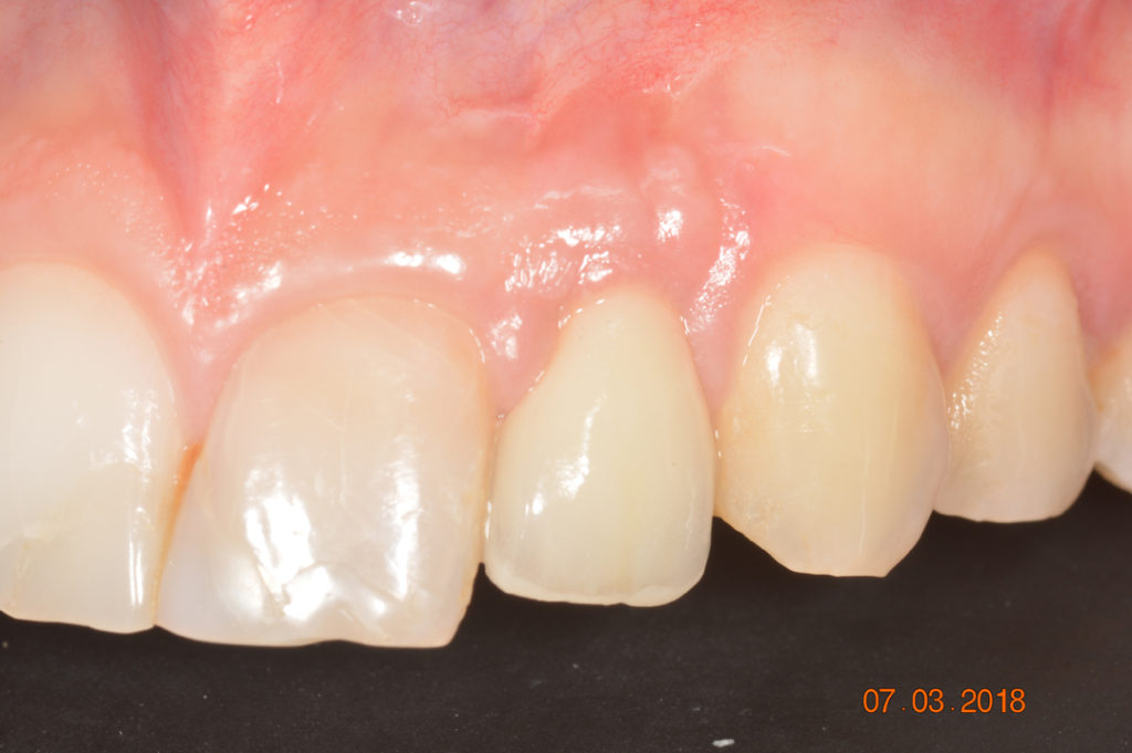
This case demonstrates the placement of a 2.9 x 14 mm XCN implant for the replacement of a congenitally missing maxillary lateral incisor of a 22-year-old female after orthodontic treatment. Immediately after placement the implant was restored with a provisional crown and a temporary abutment. After 7 months of healing, an implant level impression was taken for the fabrication of a zirconia crown on a prefabricated cylinder abutment. 1-week clinical follow-up after delivery of the final crown shows stable and healthy peri-implant tissues; an ideal interproximal papilla height has been created.

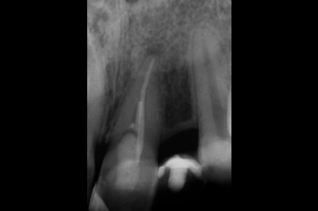
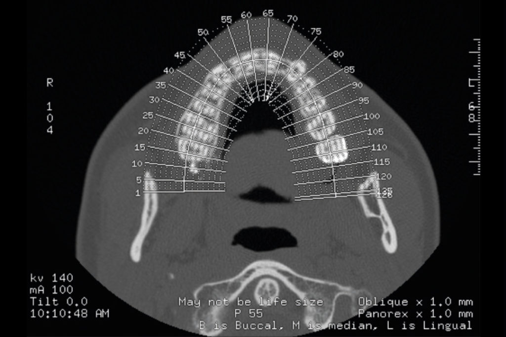
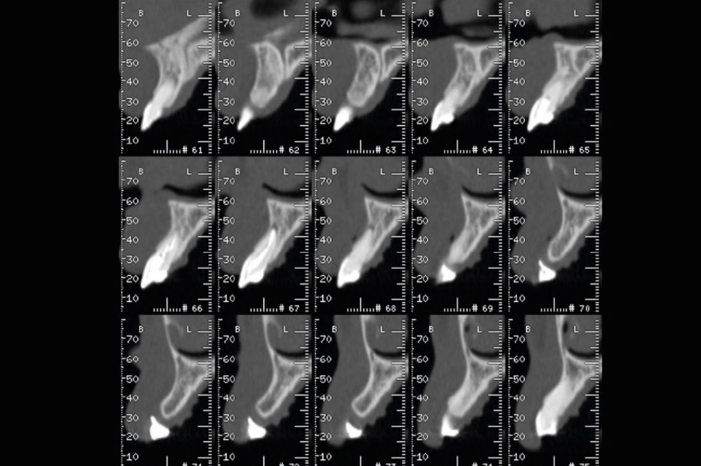
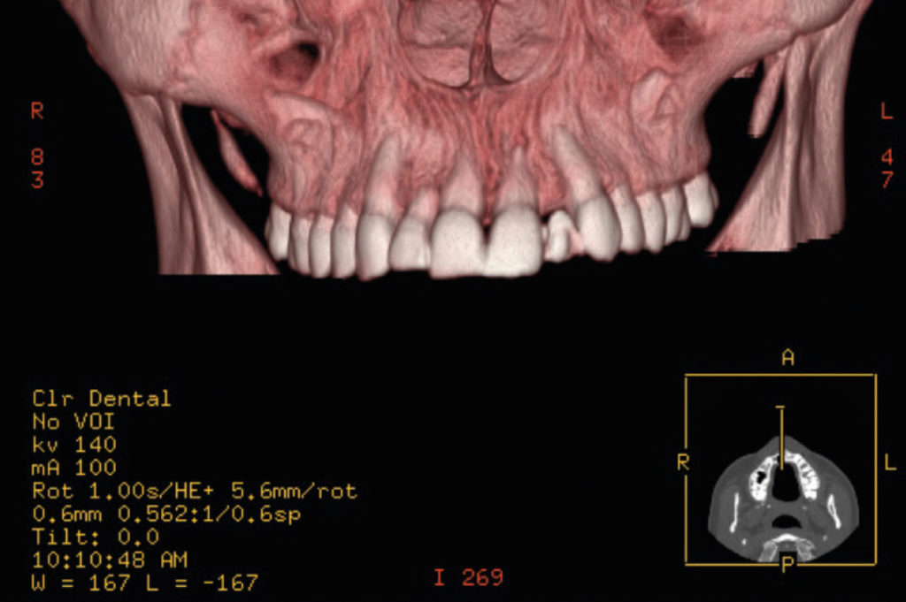
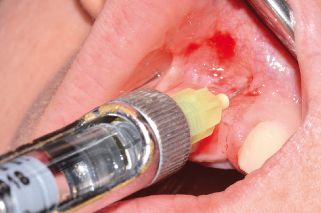
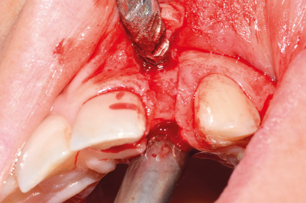
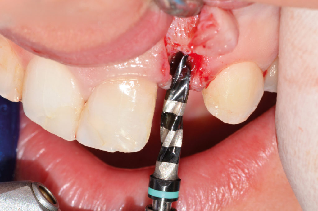
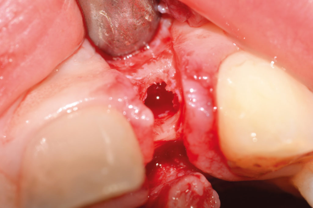
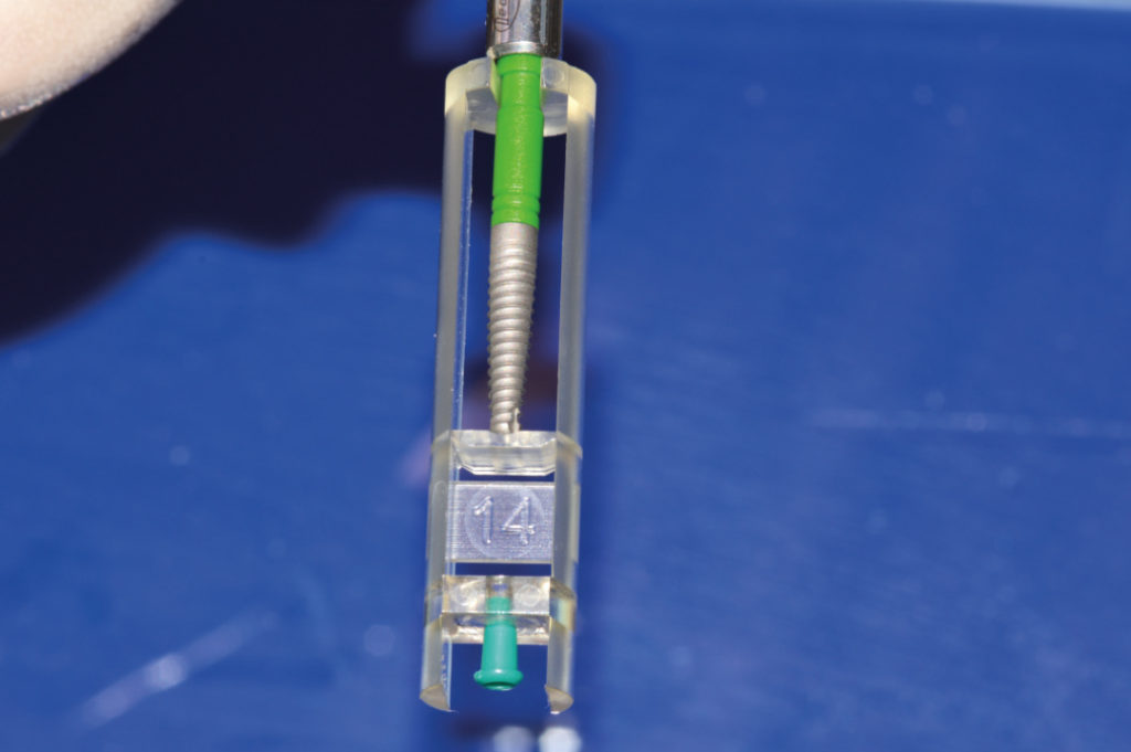
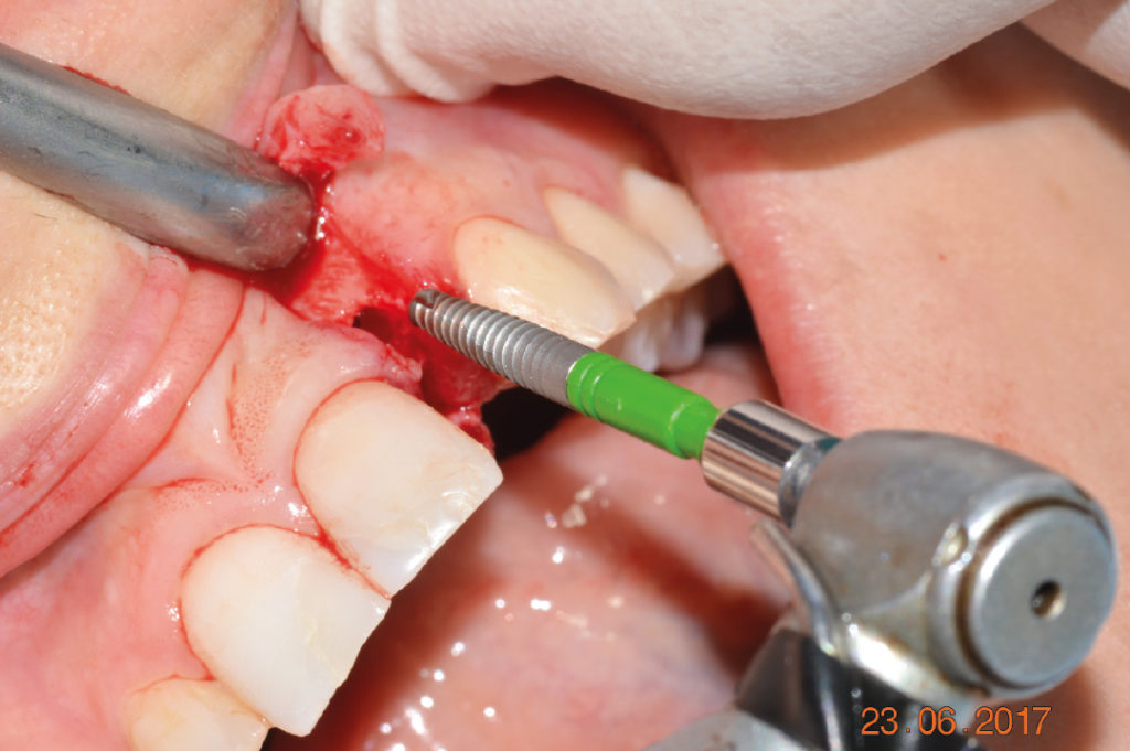
View of congenitally missing maxillary lateral incisor View of congenitally missing maxillary lateral incisor Pre-operative X-ray shows sufficient bone height for implant placement and limited interdental space Axial view with CBCT cross-section cuts CBCT cross-section images 3D reconstruction Administration of local anesthesia Rectangular full thickness flap to expose the area leaving papilla intact Creation of access hole to the bone ridge with lance drill Lance drill seated in the access hole to take an X-ray X-ray to confirm proper distance to adjacent teeth Use of 2.2 mm pilot drill for about 16 mm Use of 2.8 mm twist drill for about 6.5 mm View of created osteotomy Holder with a 2.9 x 14 mm XCN implant coupled with its carrier and cover cap Implant placement with contra-angle handpiece Implant placement with contra-angle handpiece Implant placement with contra-angle handpiece Implant seated within the osteotomy; use of a ratchet to finalize implant seating Implant placed about 2 mm subcrestally Try-in of a 3.3 mm temporary abutment Facial view of temporary restoration Lateral view of temporary restoration De-epithelization to perform a kind of roll-flap for buccal soft tissue augmentation De-epithelization to perform a kind of roll-flap for buccal soft tissue augmentation Try-in of temporary restoration Placement of vertical mattress sutures Clinical view of temporary restoration immediately after surgery Patient’s smile X-ray immediately after surgery to confirm correct implant positioning Patient’s smile two weeks after surgery Clinical view of the implant with healthy soft tissue and no recession two and a half months after implant placement X-ray two and a half months after implant placement Clinical image two weeks after surgery Clinical image one week after delivery of definitive zirconia crown 1-week clinical follow-up after crown delivery shows healthy and stable peri-implant tissues; an ideal interproximal papilla height has been created.