Surgeon/Restorative dentist:
Dr. Salvatore Belcastro – Gubbio (Perugia), Italy
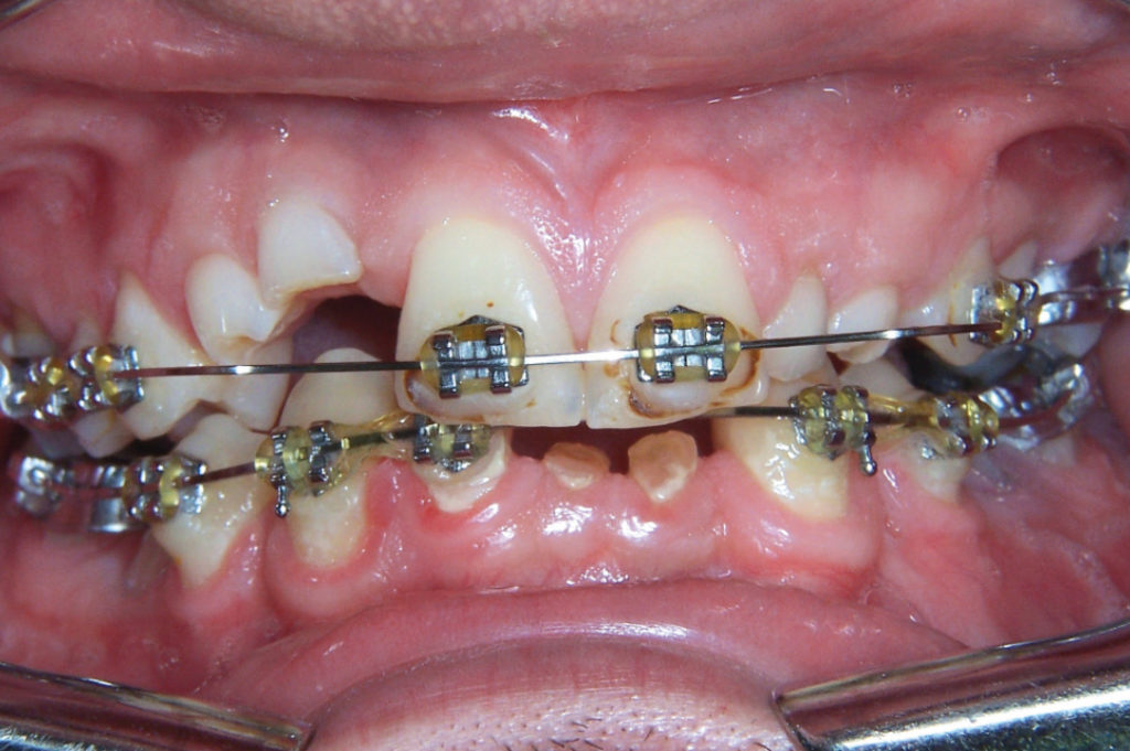
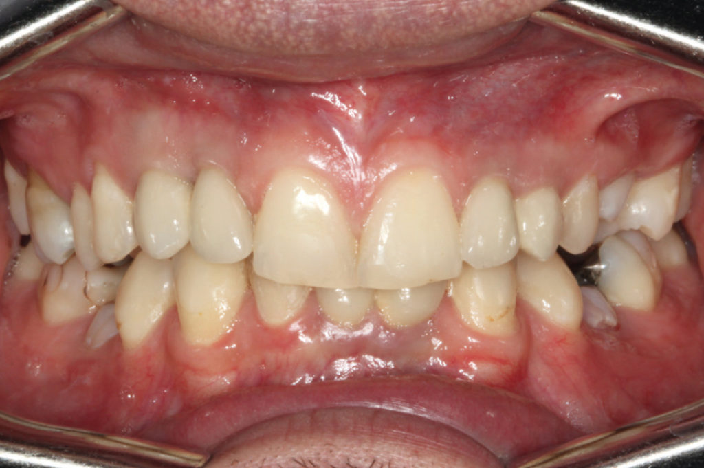
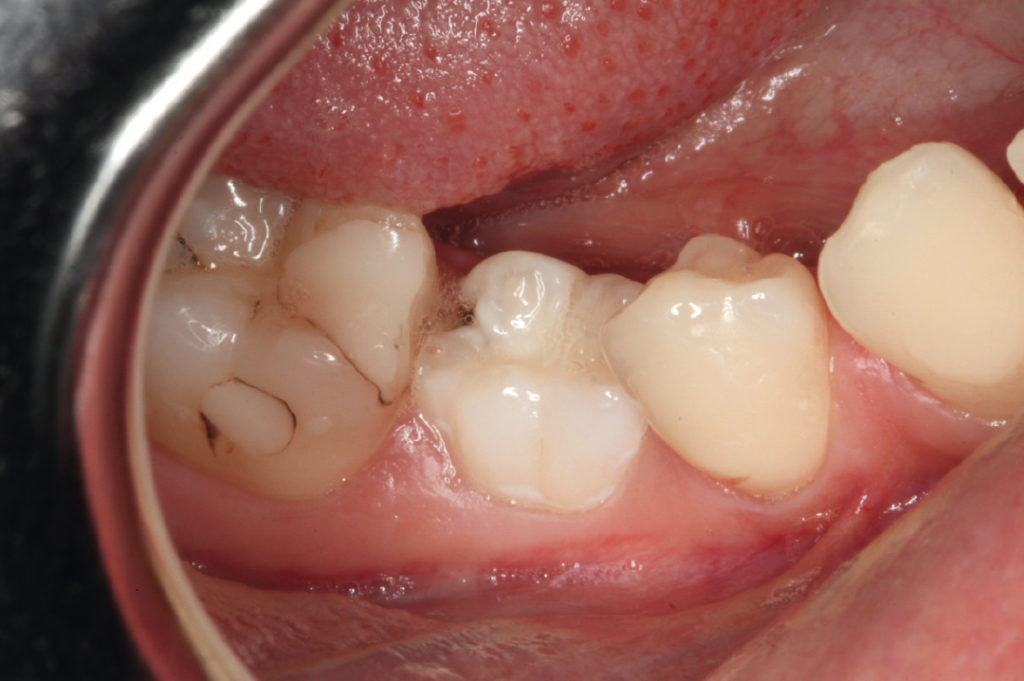
This case report demonstrates the implant treatment of several congenitally missing teeth for an eighteen-year-old male and its long-term follow-up. In total ten teeth were missing: #13, #12, #22, #23, #25, #35, #32, #31, #41 and #45. Before implant placement an orthodontic treatment was carried out for the re-distribution of the space. The implant treatment was carried out in two steps. Six implants were placed and restored in the anterior maxillary and anterior mandibular in 2009. After about one year further three implants were placed, one in maxillary left posterior region after sinus lift and two short implants replacing tooth #35 and tooth #45. Clinical and radiographic examinations were performed in July 2017 to evaluate the soft tissue and crestal bone level around implants showing stable peri-implant bone levels, healthy and intact peri-implant mucosa and excellent maintenance of aesthetic results.

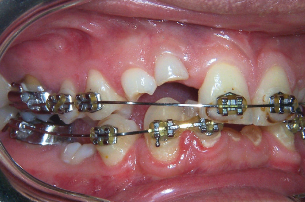
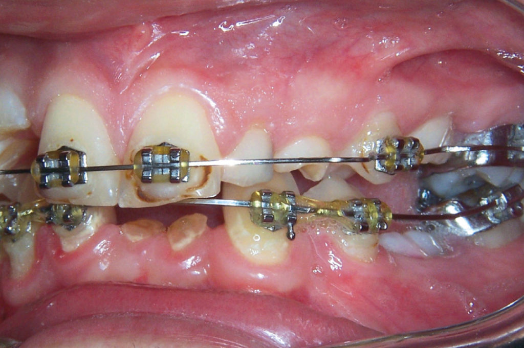
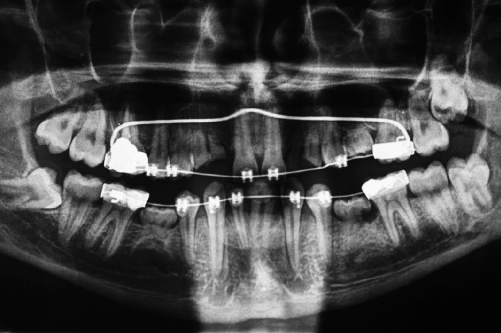
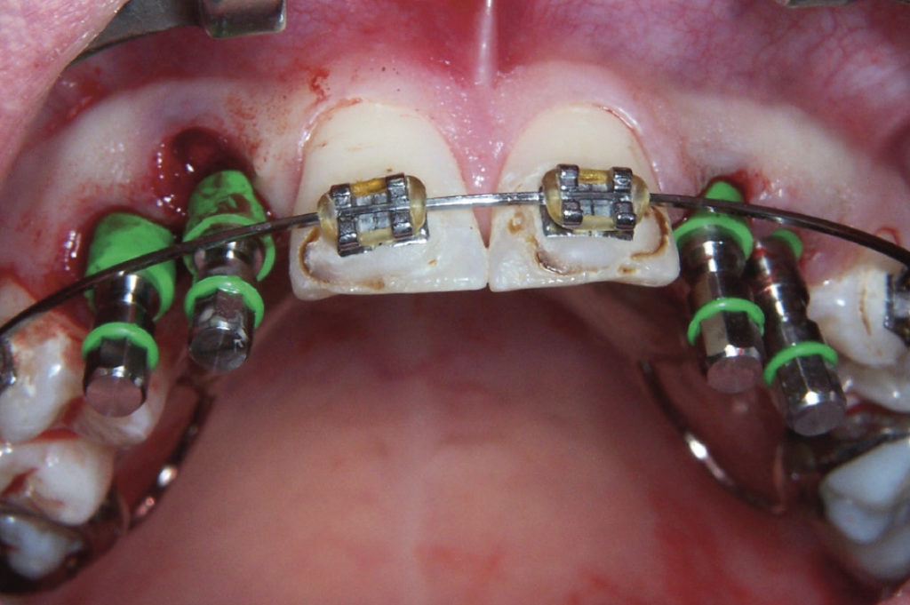
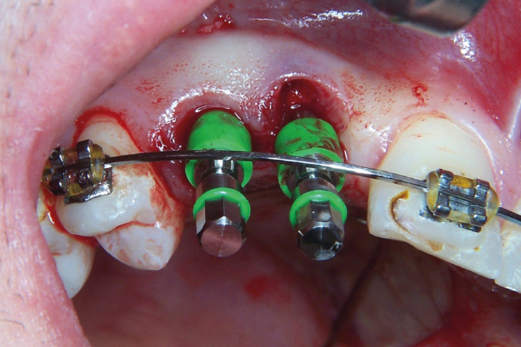
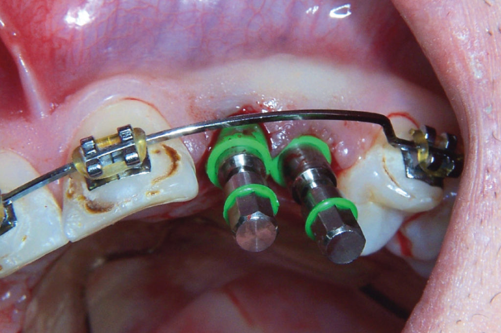
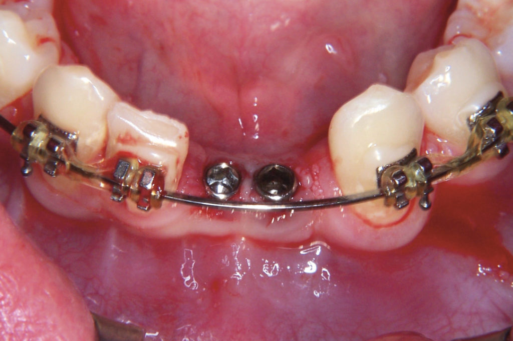
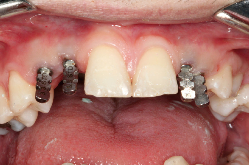
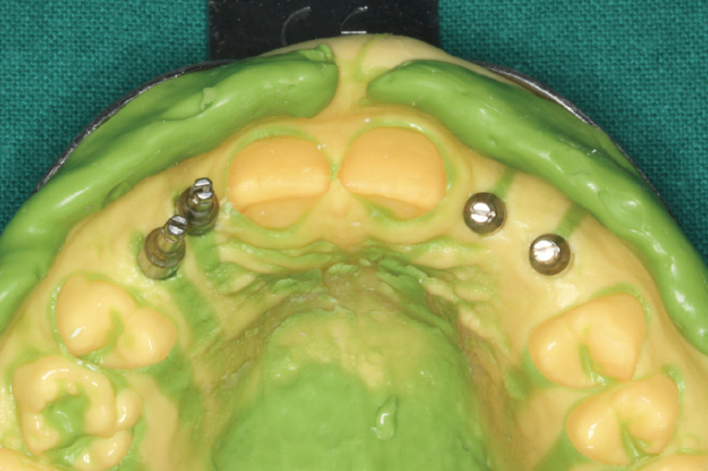
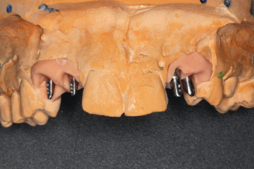
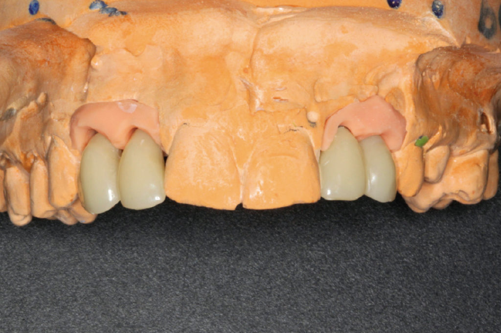
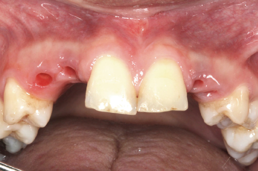
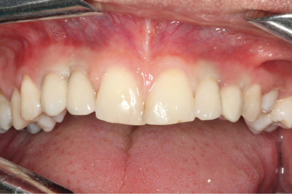
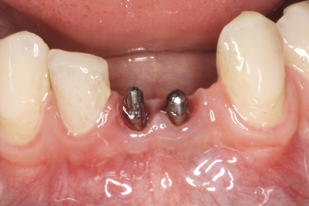
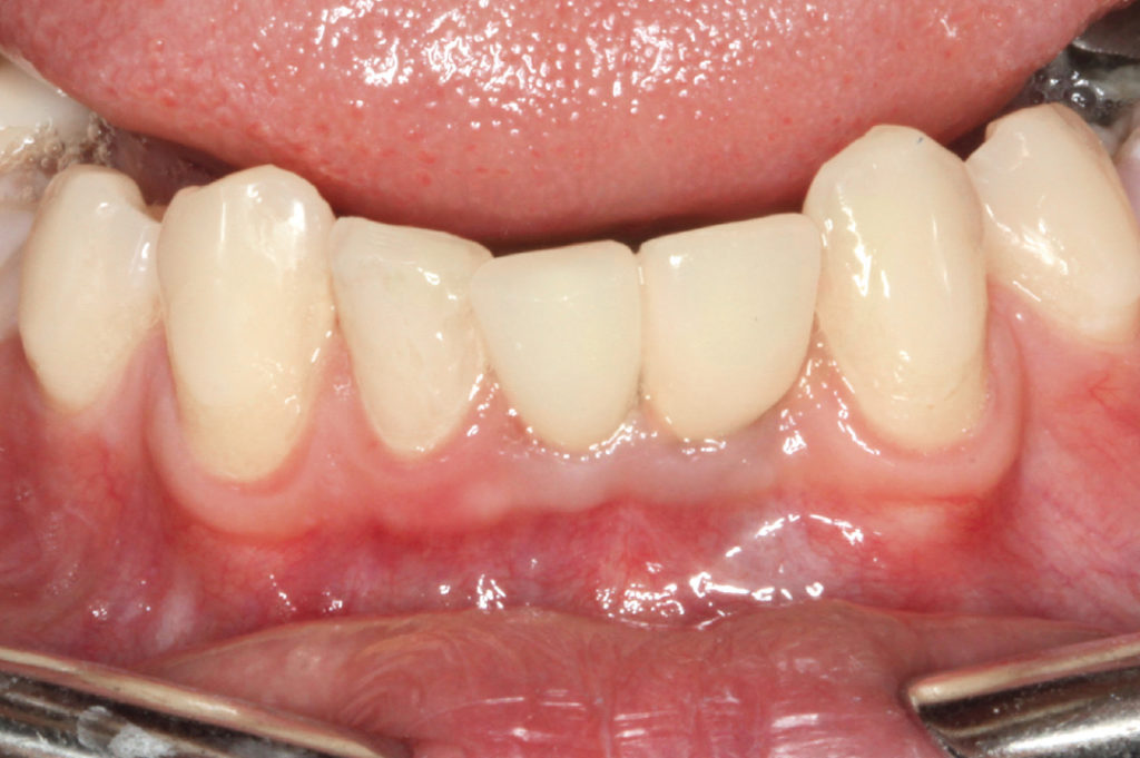
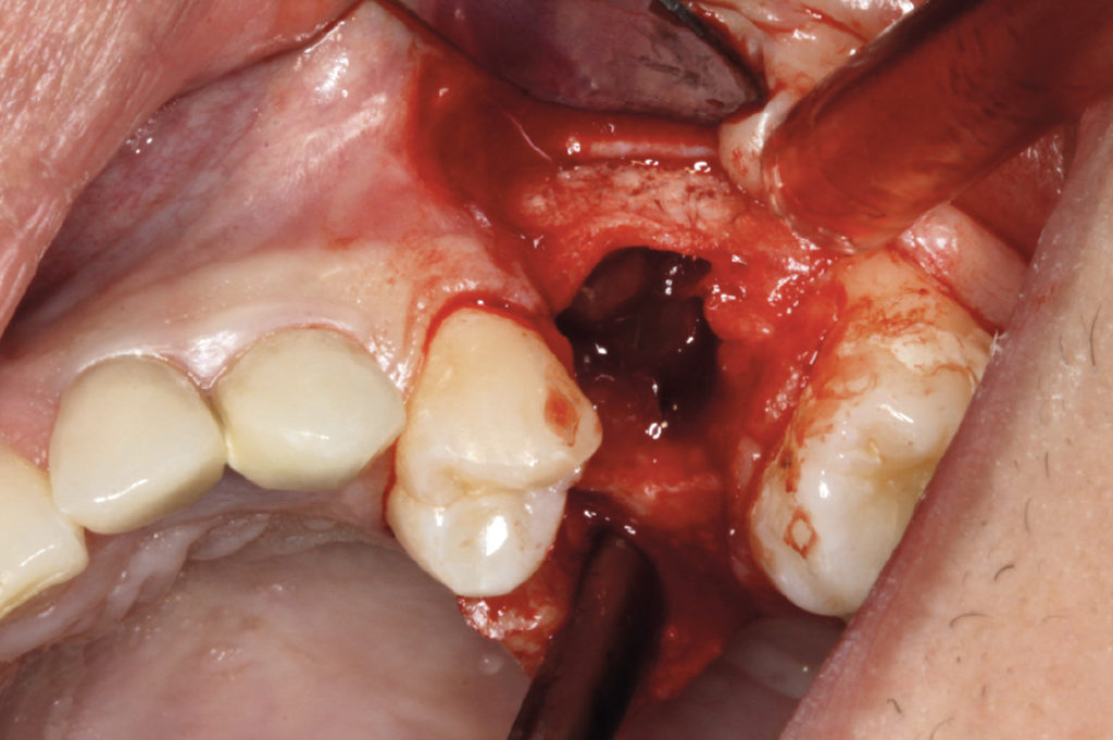
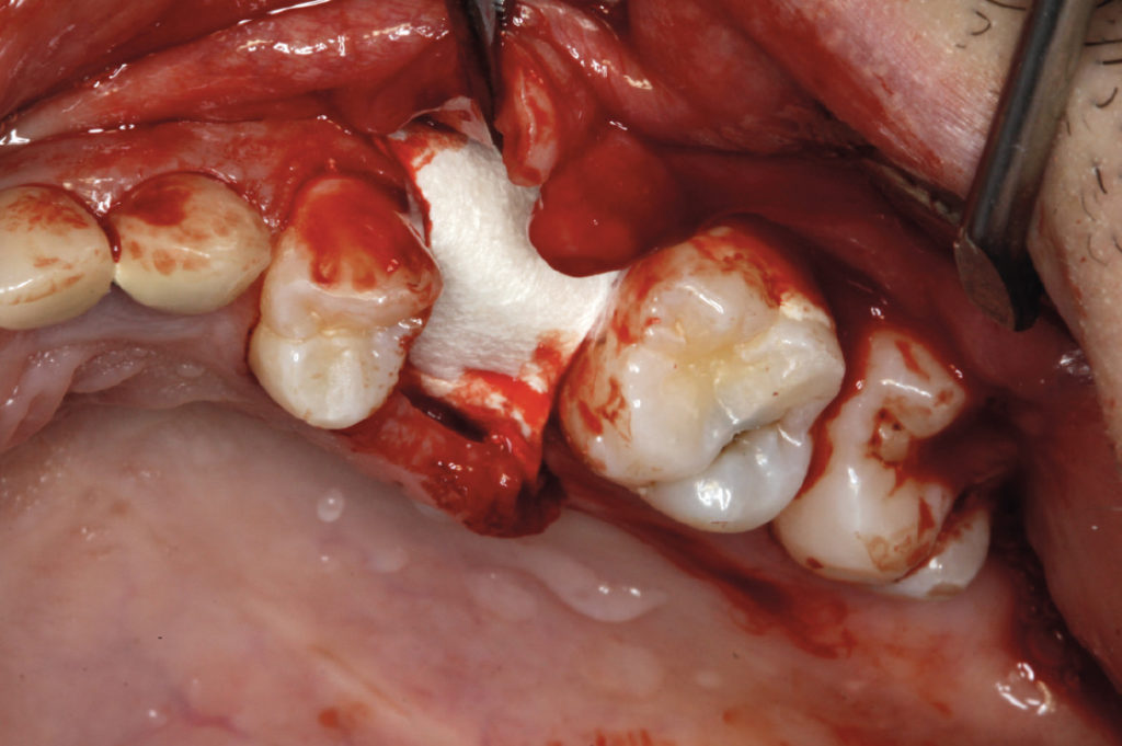
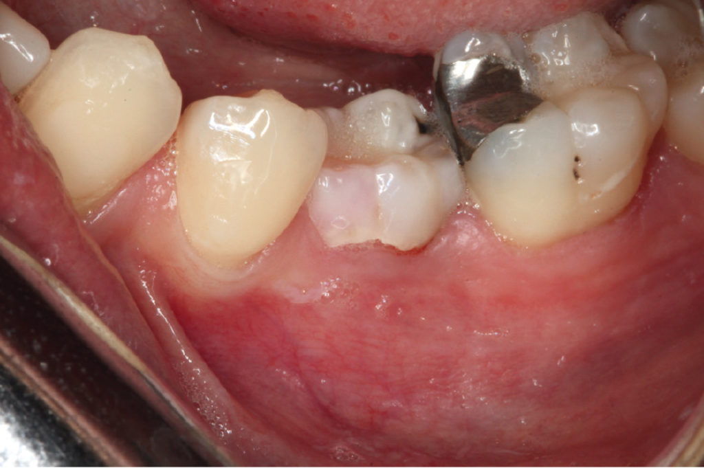
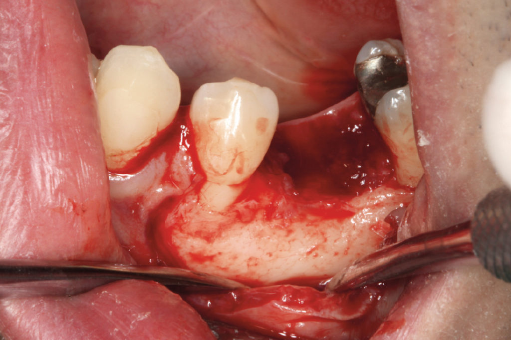
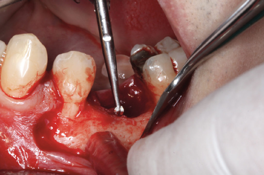
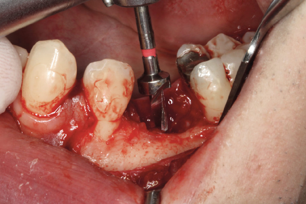
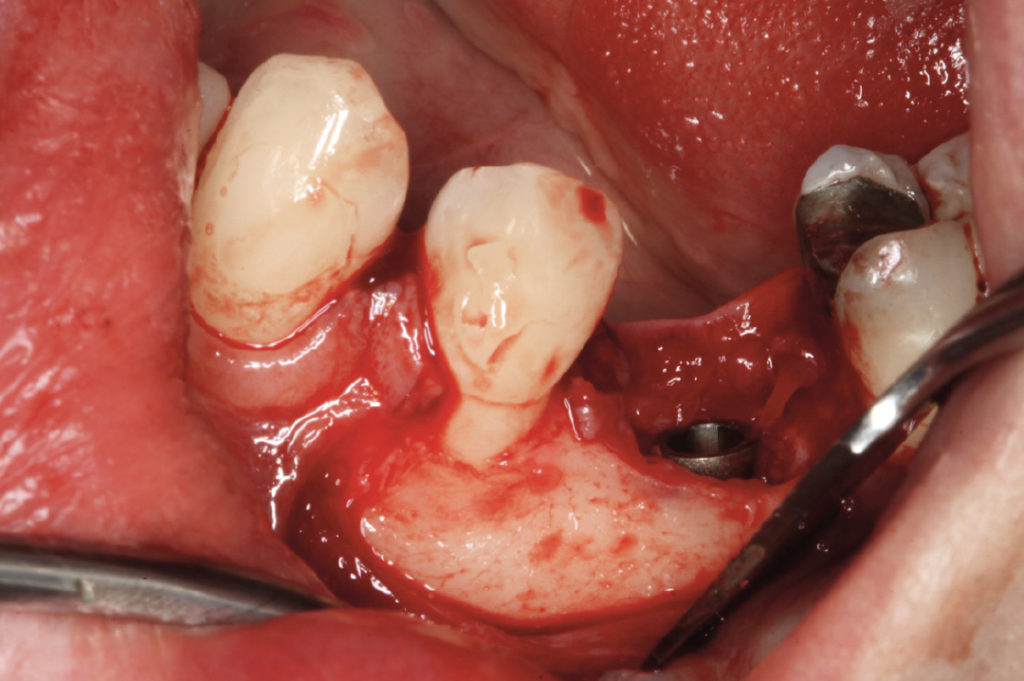
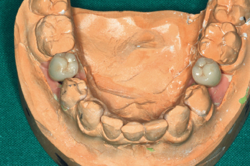
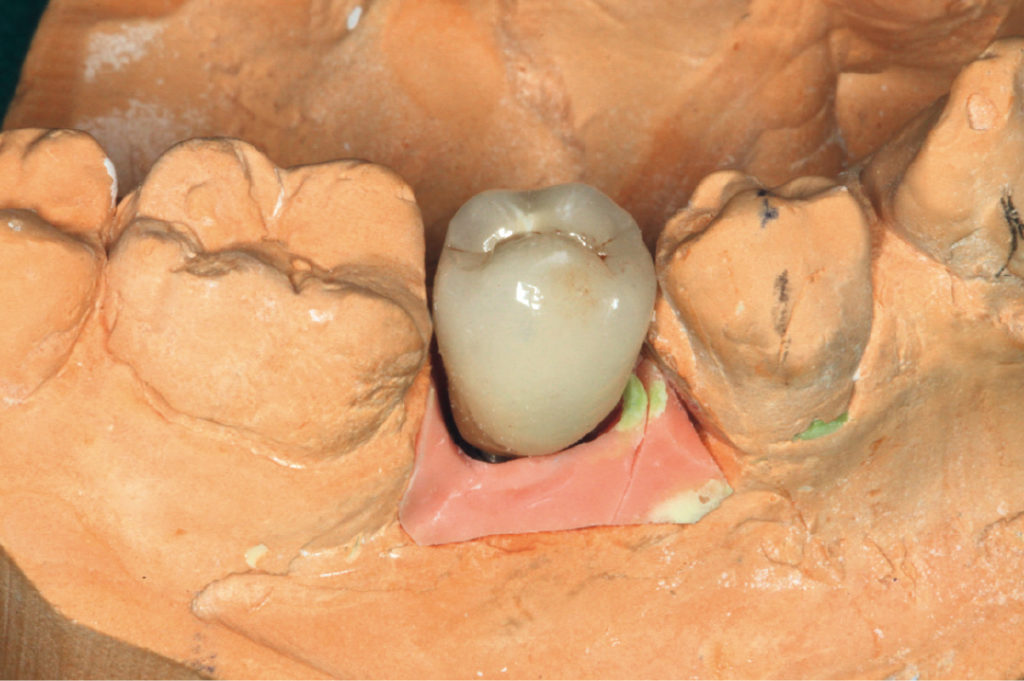
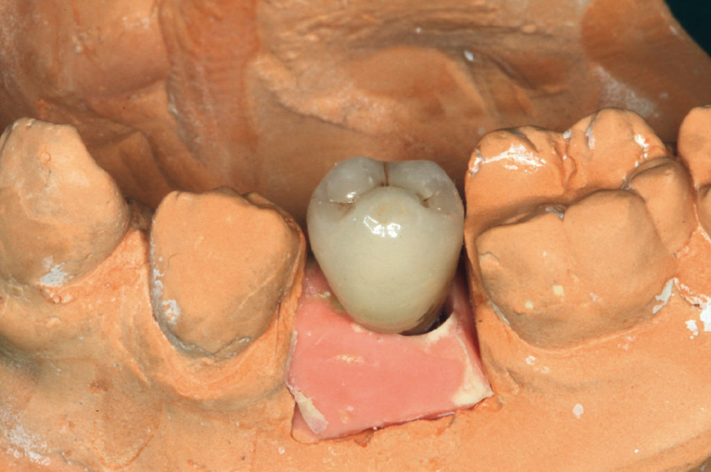
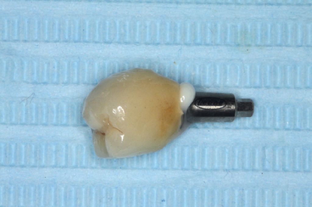
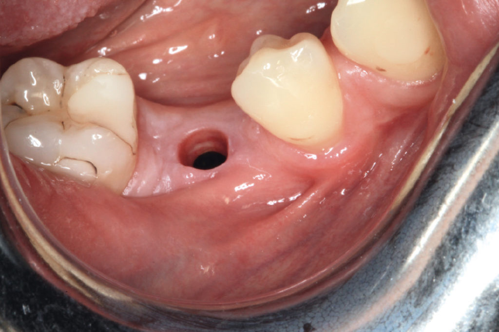
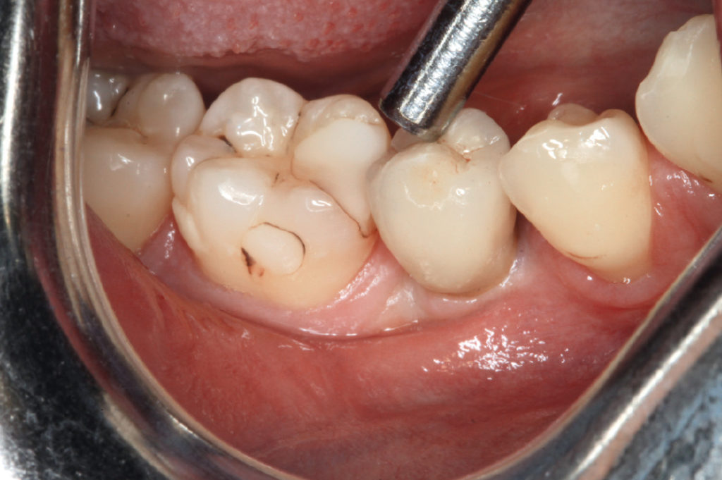
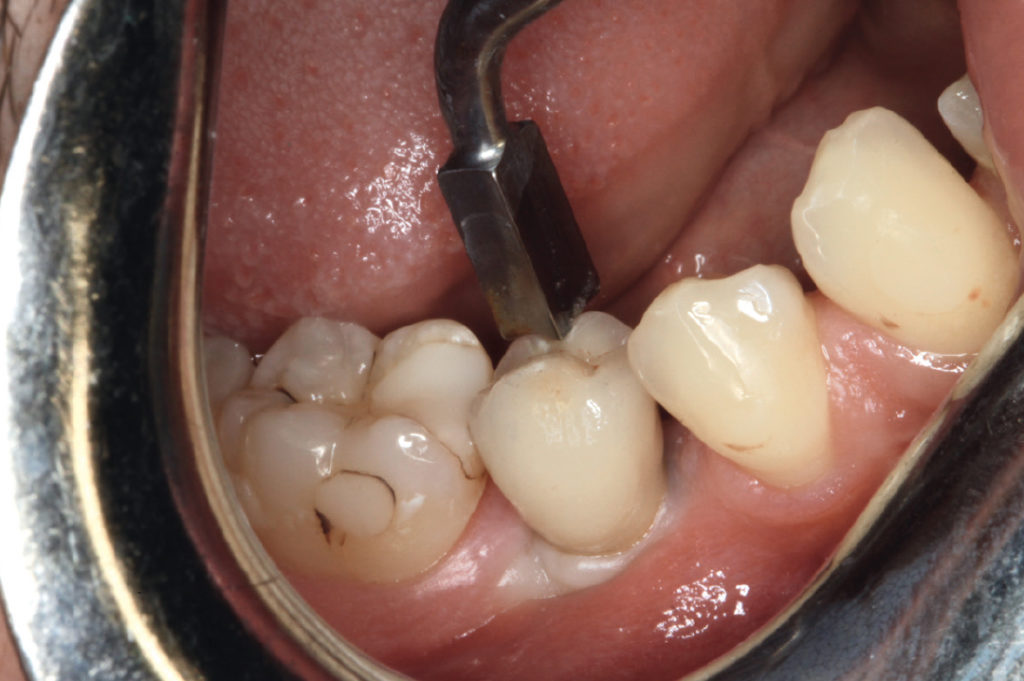
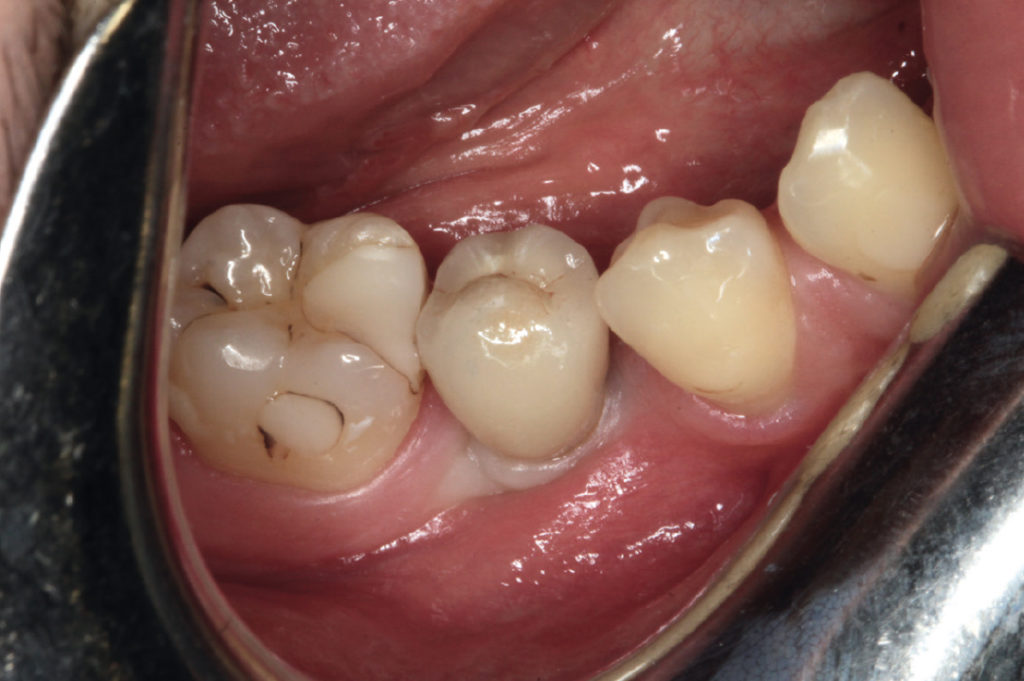
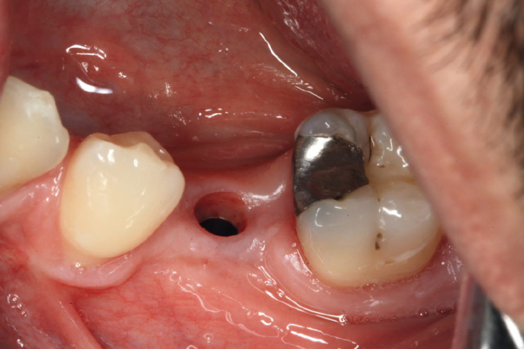
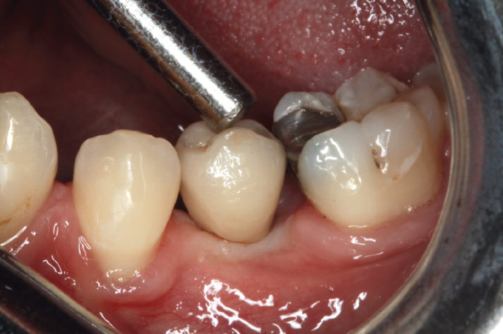
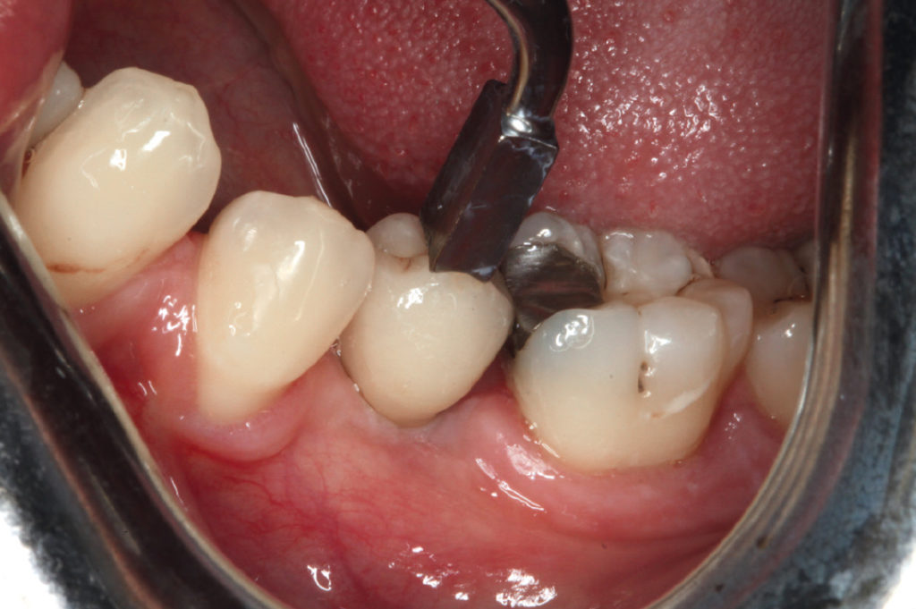
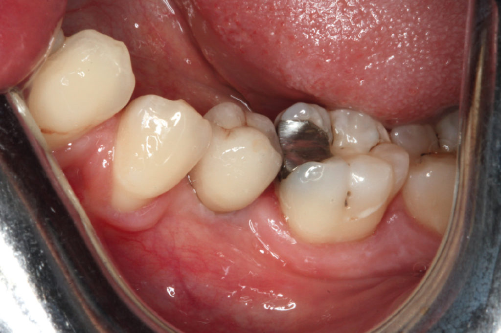
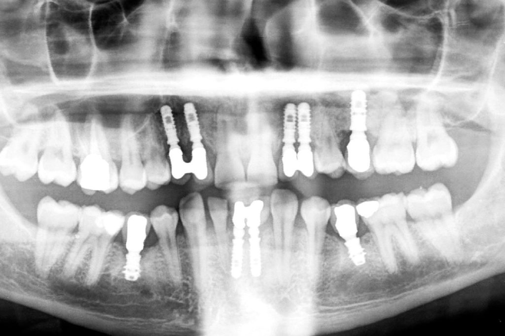
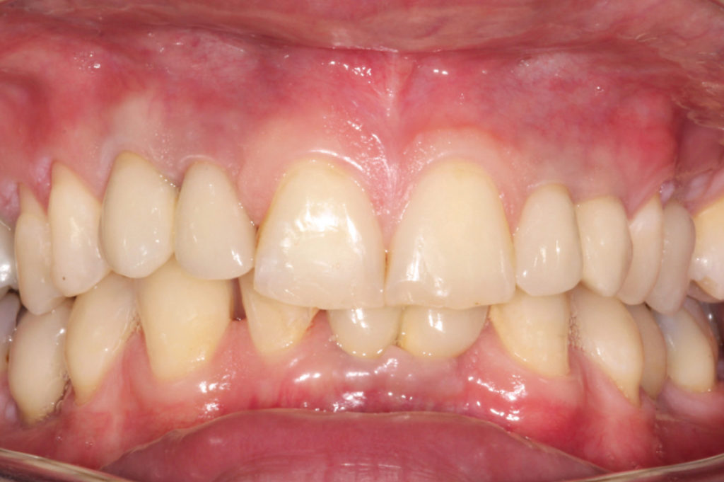
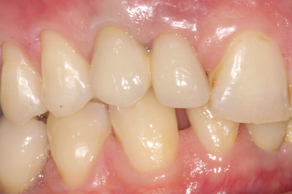
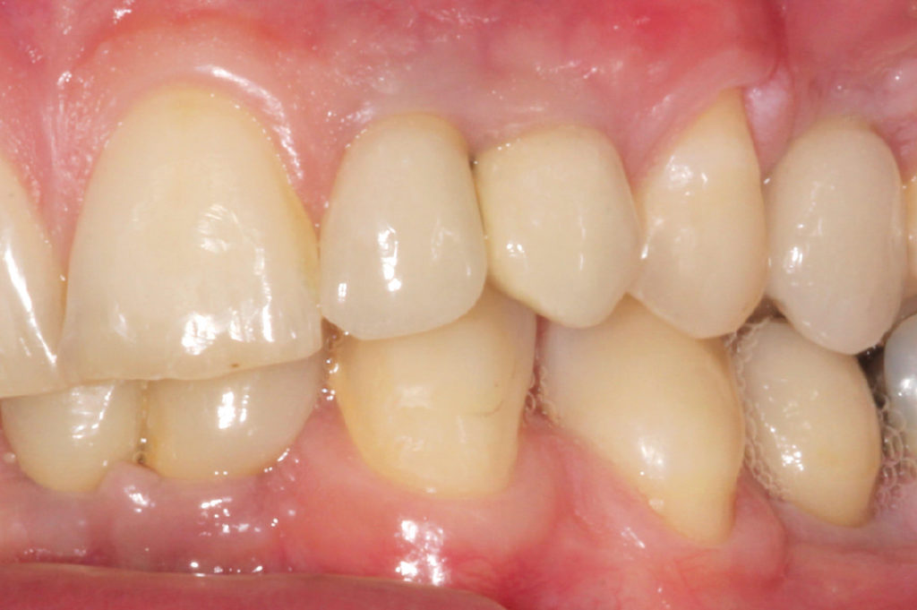
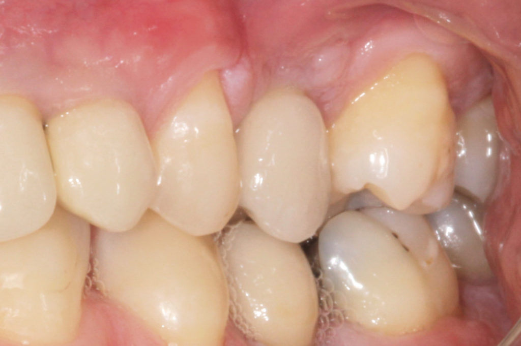
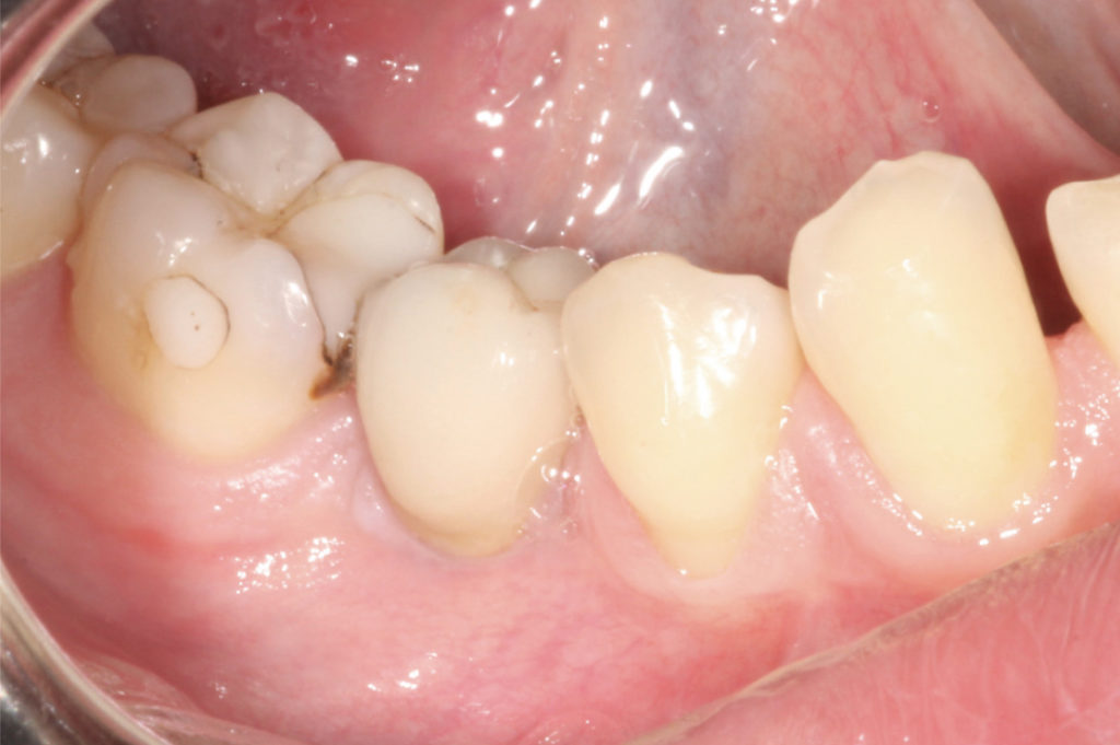
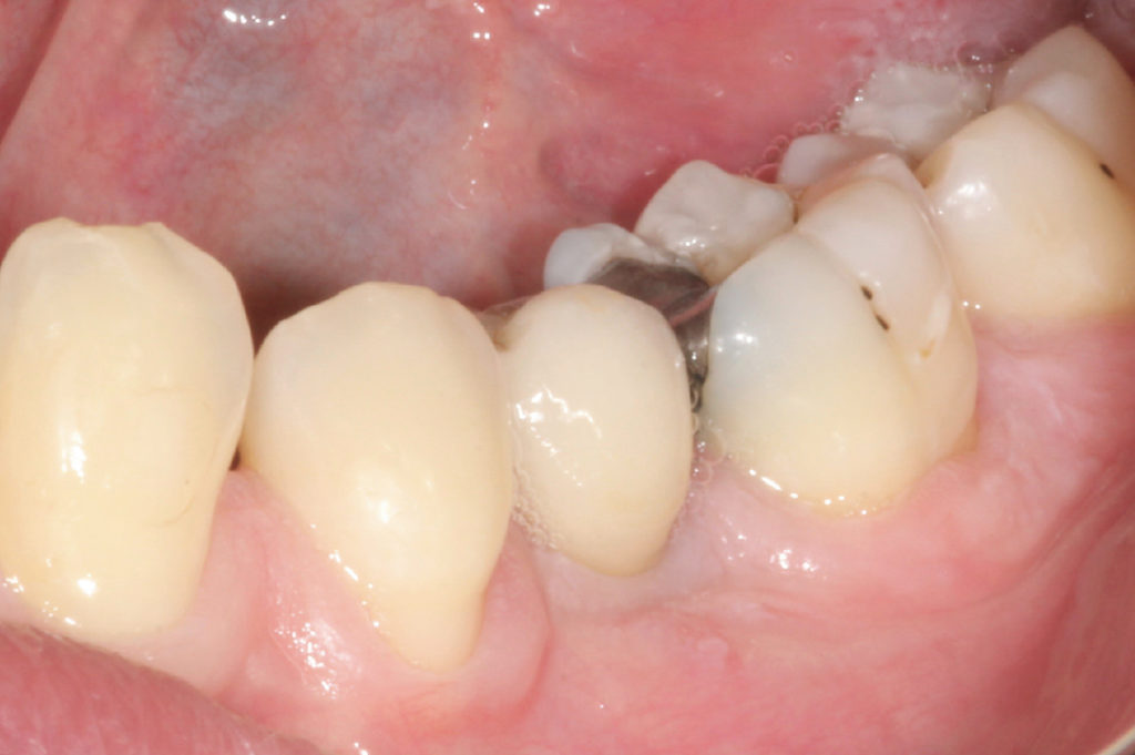
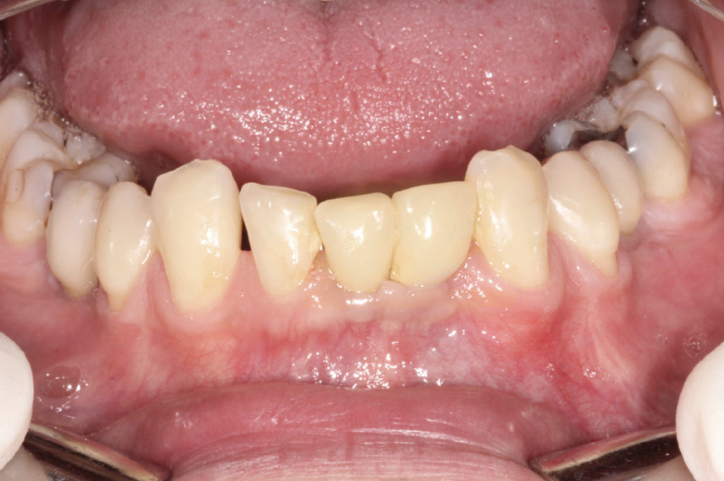
Clinical situation after orthodontic treatment View of right upper and lower quadrants after orthodontic treatment View of left upper and lower quadrants after orthodontic treatment Panoramic X-ray after orthodontic treatment In 2009: placement of four 3.3 x 10 mm XCN Classix implants in the upper jaw View of the carrier of two 3.3 x 10 mm XCN Classix implants in the right upper jaw View of the carrier of two 3.3 x 10 mm XCN Classix implants in the left upper jaw In 2009: placement of two 3.3 x 10 mm XCN Classix implants in the anterior lower jaw replacing teeth #32, #31 and #41 Four months post-placement view of transfers positioned in the anterior upper implants for impression taking Transfers inside the impression Try-in of prepared abutments on the dental cast Try-in of abutments and metal ceramic restorations on the dental cast View of healthy soft tissues prior to incorporating the definitive restorations Delivery of the definitive restorations in the anterior upper jaw Intraoral try-in of prepared titanium abutments in the anterior lower jaw Delivery of the definitive restorations in the anterior lower jaw Six months post-insertion clinical view In 2010: view of sinus lift in maxillary left posterior region for replacing tooth #25 Covering the access to the maxillary sinus with resorbable collagen membrane In 2010: deciduous tooth #85 In 2010: deciduous tooth #75 Full thickness flap to expose the bone in the region of tooth #35 Use of round bur to mark the cortical bone Implant site preparation for the placement of a Leone 6.5 short implant Leone 6.5 short implant in place Cover cap positioned on the implant Four months later: definitive restorations on the dental cast Try-in of abutment and metal ceramic crown for tooth #45 on the dental cast Try-in of abutment and metal ceramic crown for tooth #35 on the dental cast Extraoral cementation of metal ceramic crown on abutment View of healthy soft tissues prior to incorporating the definitive restoration of tooth #45 Intraoral try-in of the restoration for tooth #45 Definitive restoration for tooth #45 is tapped into the implant Clinical view of tooth #45 immediately after incorporating the definitive restoration View of healthy soft tissues prior to incorporating the definitive restoration of tooth #35 Intraoral try-in of the restoration for tooth #35 Definitive restoration for tooth #35 is tapped into the implant Clinical view of tooth #35 immediately after incorporating the definitive restoration Panoramic X-ray in July 2017. Note excellent crestal bone level maintenance after eight resp. seven years Clinical situation in July 2017. Note healthy and intact peri-implant mucosa View of teeth #13 and #12 eight years post insertion View of teeth #22 and #23 eight years post insertion View of tooth #25 seven years post insertion View of tooth #45 seven years post insertion View of tooth #35 seven years post insertion View of anterior lower jaw eight years post insertion

Laboratory:
Wilocs – Rome, Italy