Surgeon/Restorative dentist:
Dr. Alberto Frezzato – Rovigo, Italy
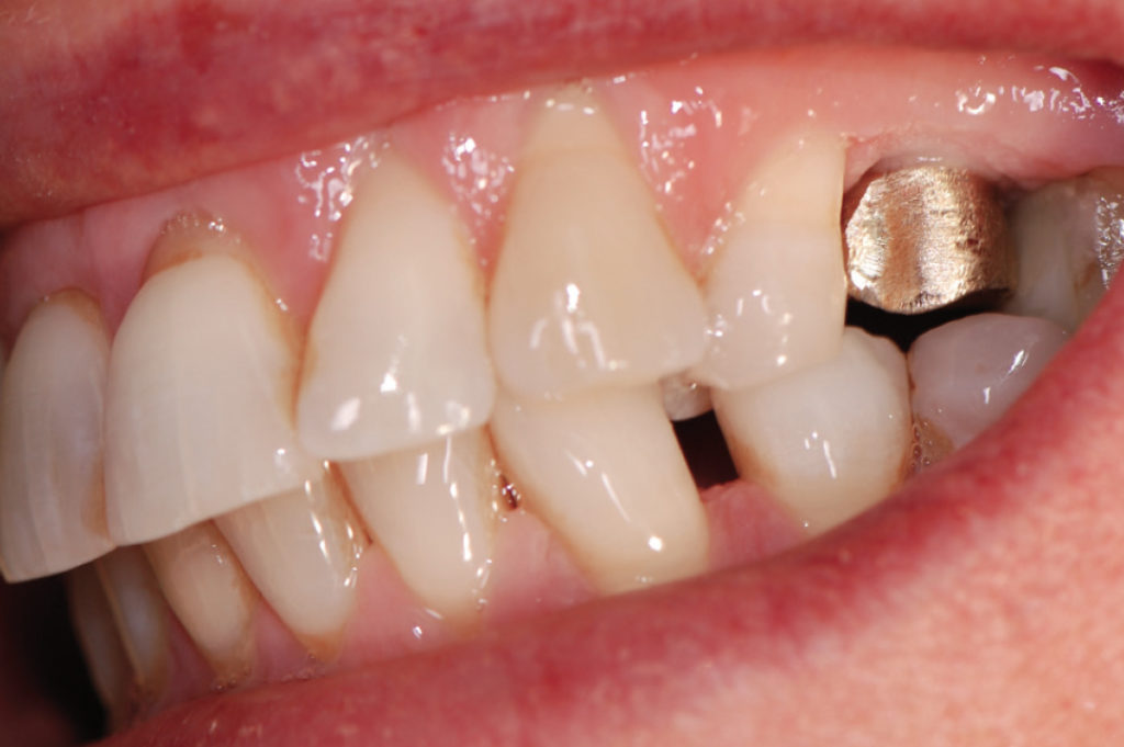
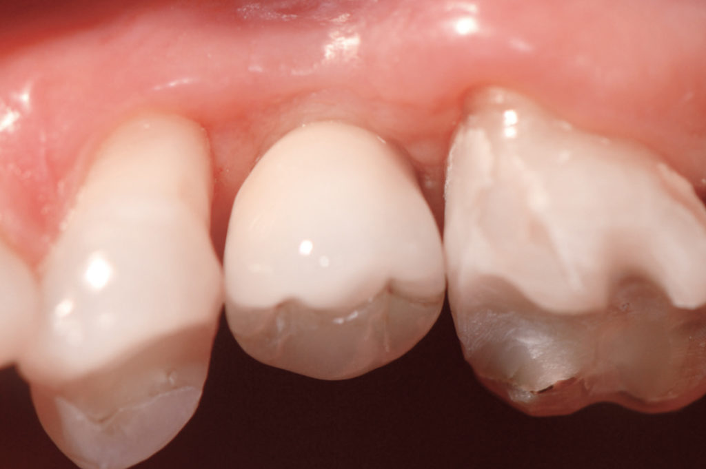
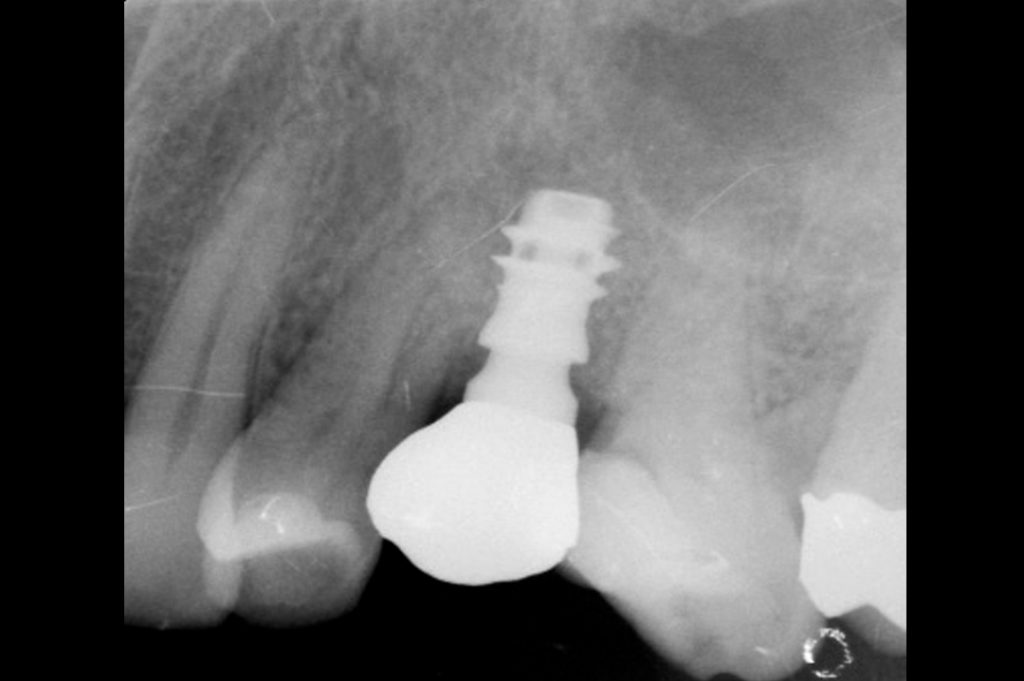
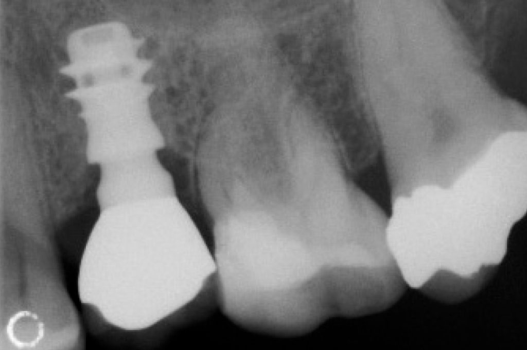
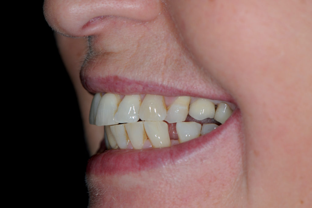
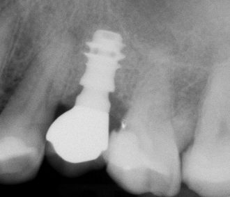
This case report illustrates immediate implant placement of an XCN 6.5 short implant for replacing tooth #25 of a 52-year-old female. Clinical and radiographic examination revealed sufficient interdental space, sufficient ridge width, thick gingival biotype and a residual bone height of only 7 mm. A minimally invasive surgery was performed: atraumatic root extraction, flapless approach, immediate placement of an implant with a length of only 6.5 mm and immediate insertion of a healing cap for a one-stage surgical procedure. After four months of healing, an implant level impression was taken for the fabrication of an extra-orally cemented metal ceramic crown. 5-year clinical and radiographic follow-up demonstrate the maintenance over time of the achieved results: stable peri-implant marginal bone levels, stable peri-implant soft tissue levels and excellent gingival tissue health. The radiographic follow-up 8 years after delivery of the definitive crown shows the same situation: no peri-implant marginal bone loss can be observed.

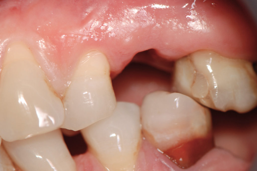
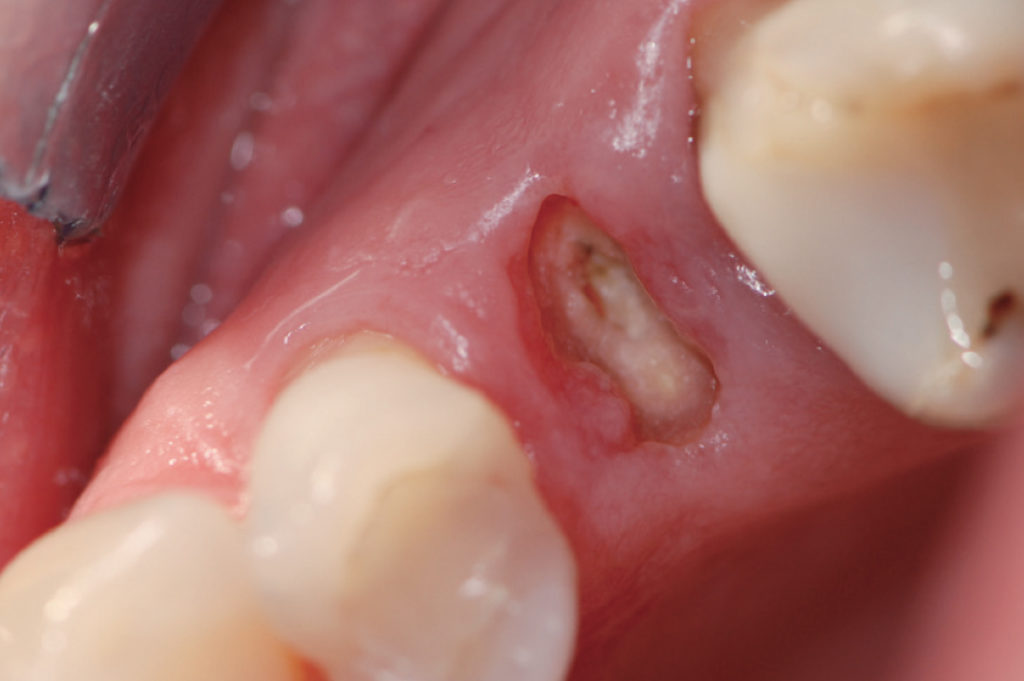
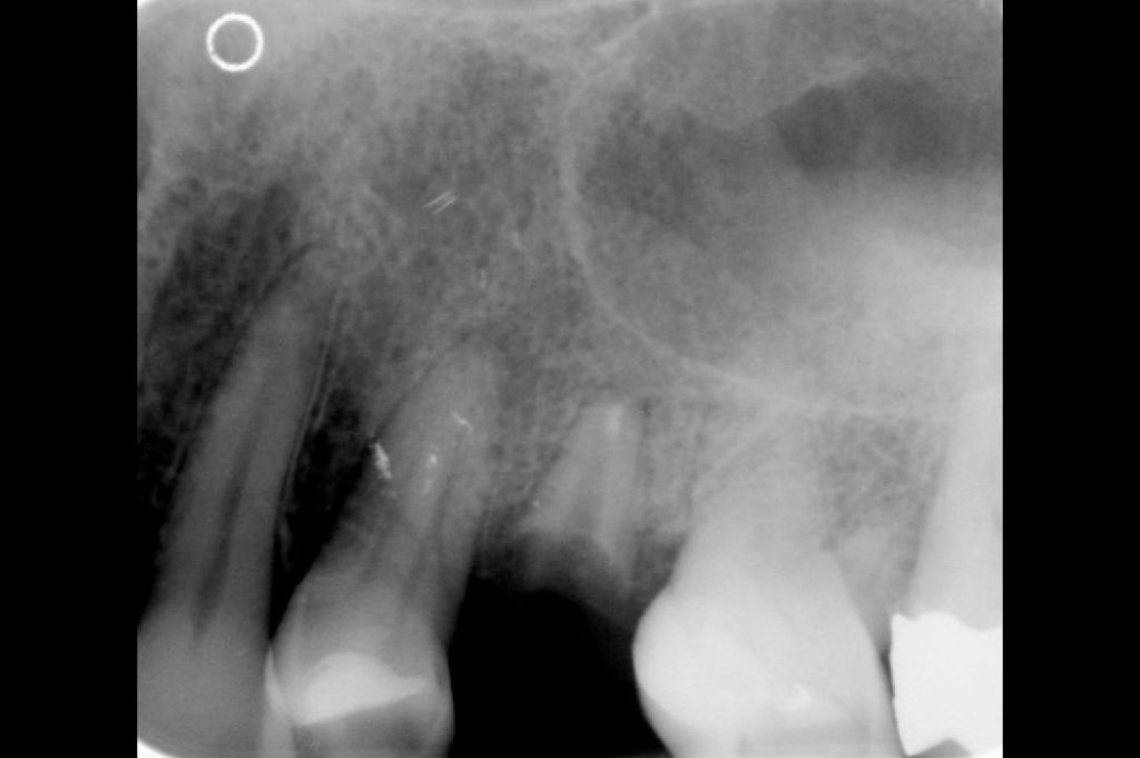
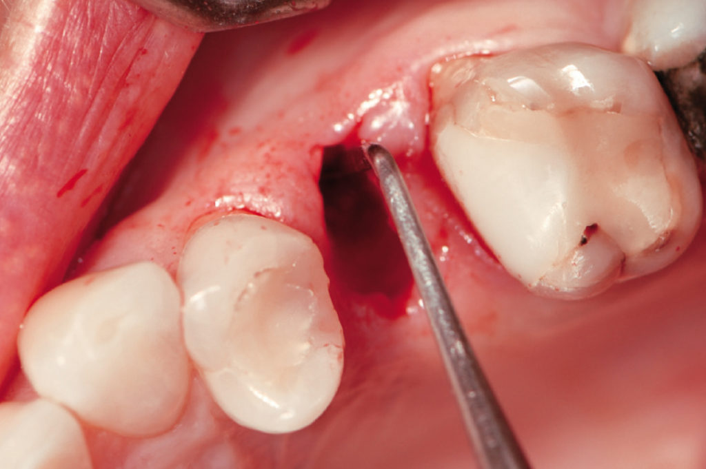
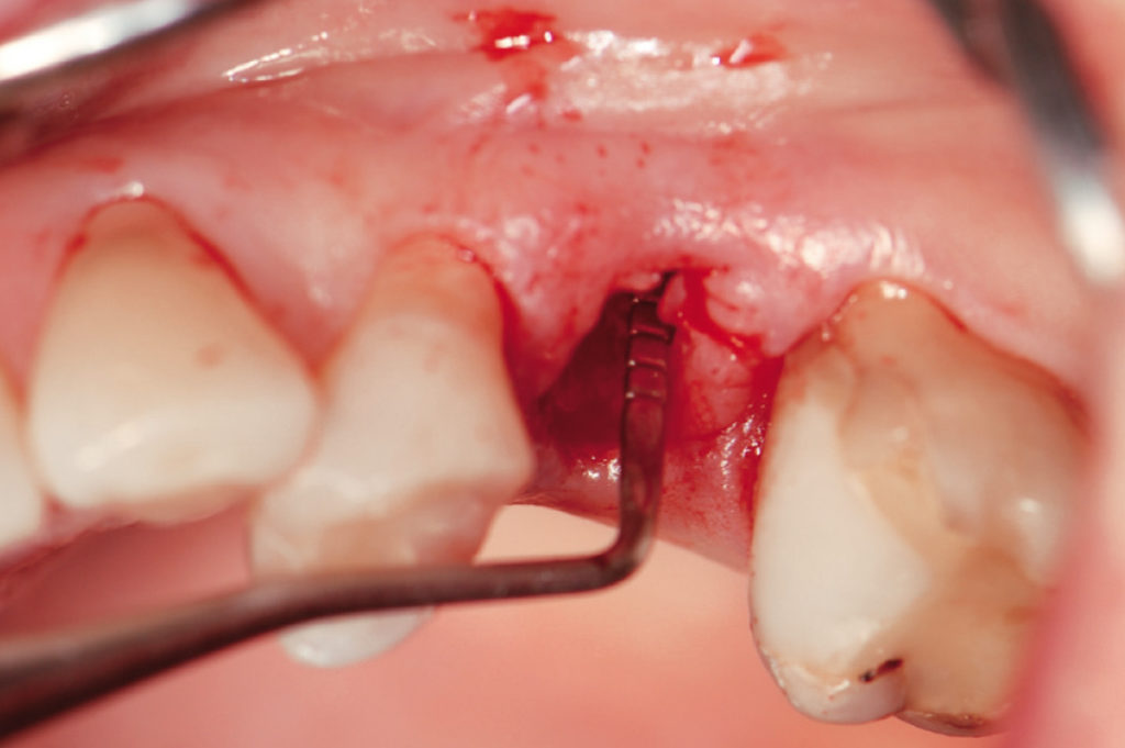
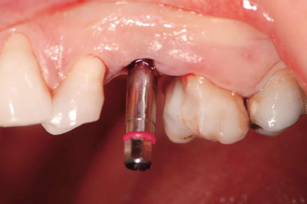
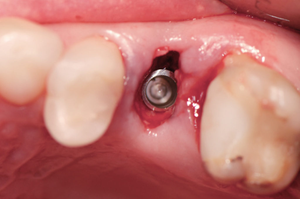
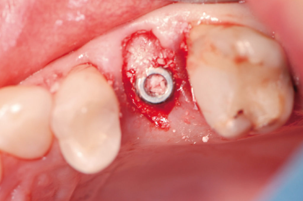
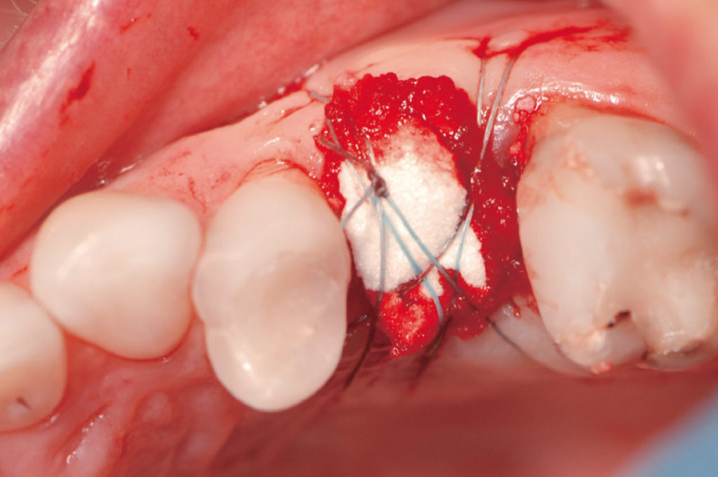
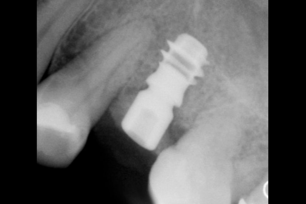
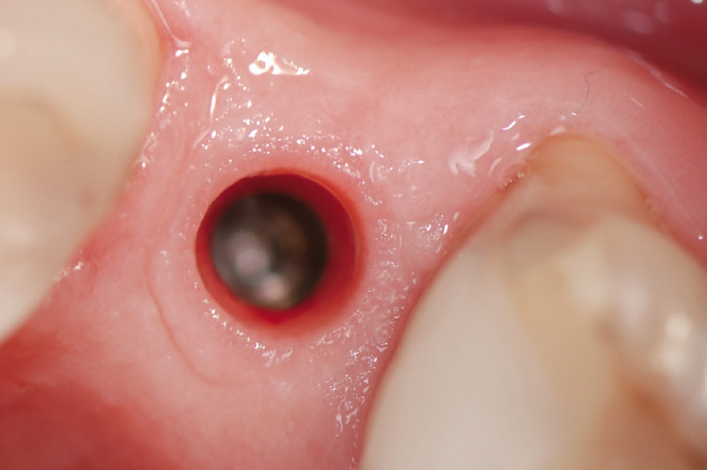
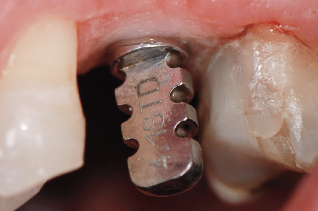
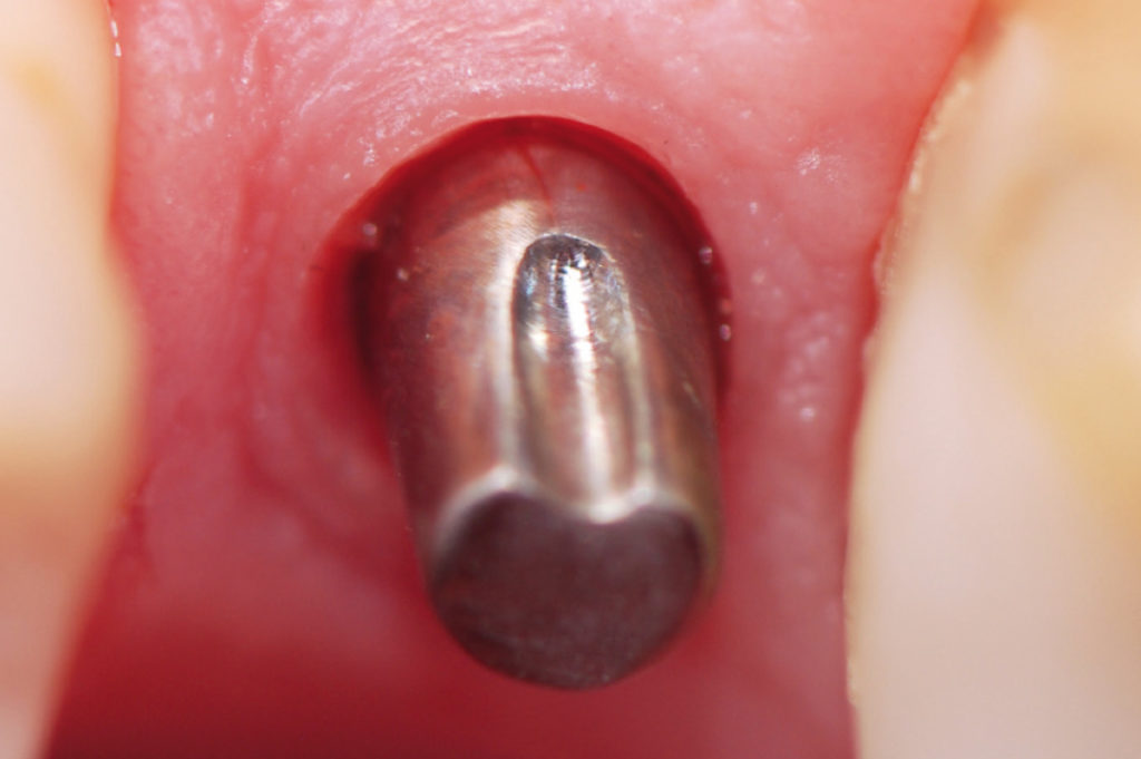
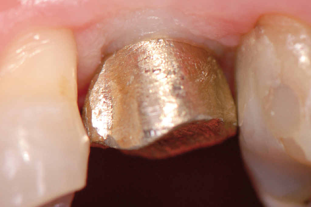
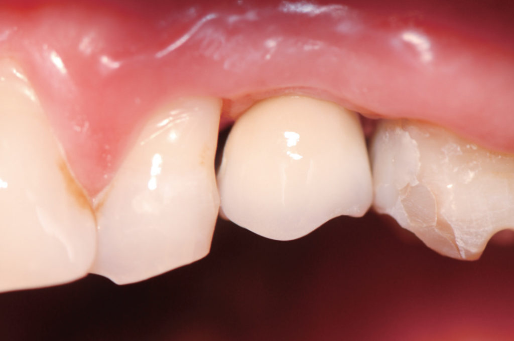
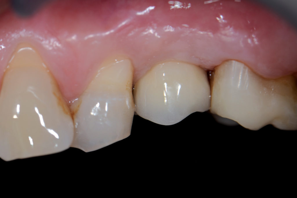
Lateral view of missing upper left second premolar Occlusal view of missing upper left second premolar Pre-operative X-ray Occlusal view immediately after flapless root extraction. Inspection of the post-extraction socket to confirm that all bone walls are intact Inspection of the buccal bone wall Preparation of the implant site for an XCN 6.5 short implant Occlusal view of the implant in place Implant closed with a healing cap GH5; gap between implant and buccal bone wall filled with synthetic bone grafting material Placement of a collagen sponge to cover the implant site Post-operative X-ray Four months post-operative clinical view View of peri-implant soft tissues after removal of the healing cap. Note excellent health of peri-implant soft tissues Transfer placed within the implant Try-in of the prepared abutment Try-in of the metal structure Try-in of the metal structure Delivery of extra-orally cemented metal ceramic crown Lateral view after crown delivery X-ray after crown delivery Clinical situation five years after crown delivery. Note excellent health and stability of peri-implant soft tissues 5-year follow-up X-ray. Note stable peri-implant crestal bone levels Patient’s smile at 5-year follow-up 8-year follow-up X-ray. No peri-implant marginal bone loss can be observed

Laboratory:
Ceramodent, Paolo Morbiato & C. – Padua, Italy
Link to the original article published in 2010
5-year follow-up article was published in 2014: Frezzato A, Frezzato I, L’impianto singolo. Dalle evidenze scientifiche ai risultati clinici, ISO 2014, p. 108-112