Surgeon/Restorative dentist:
Dr. Francesco Argentino – Florence, Italy
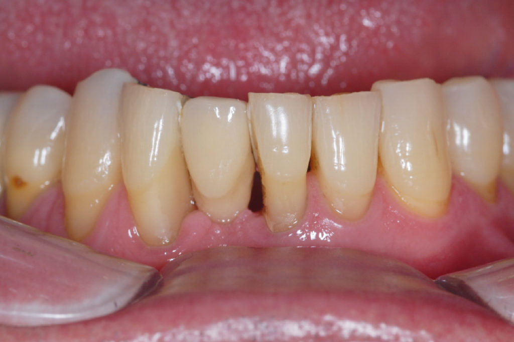
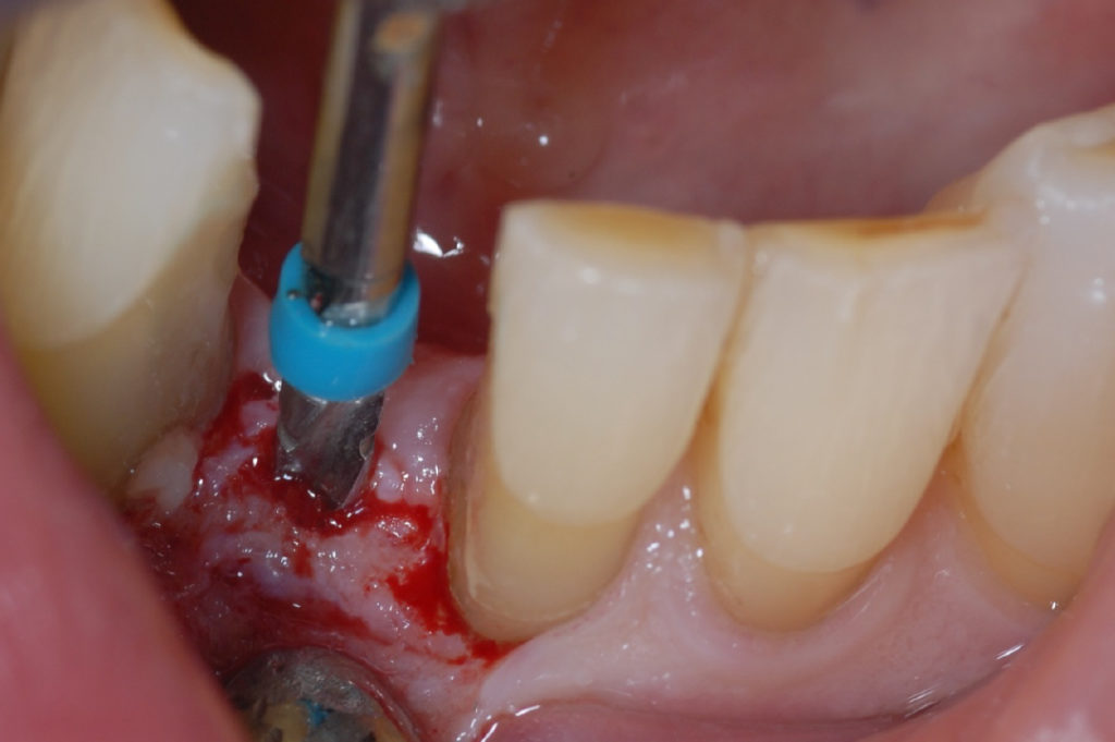
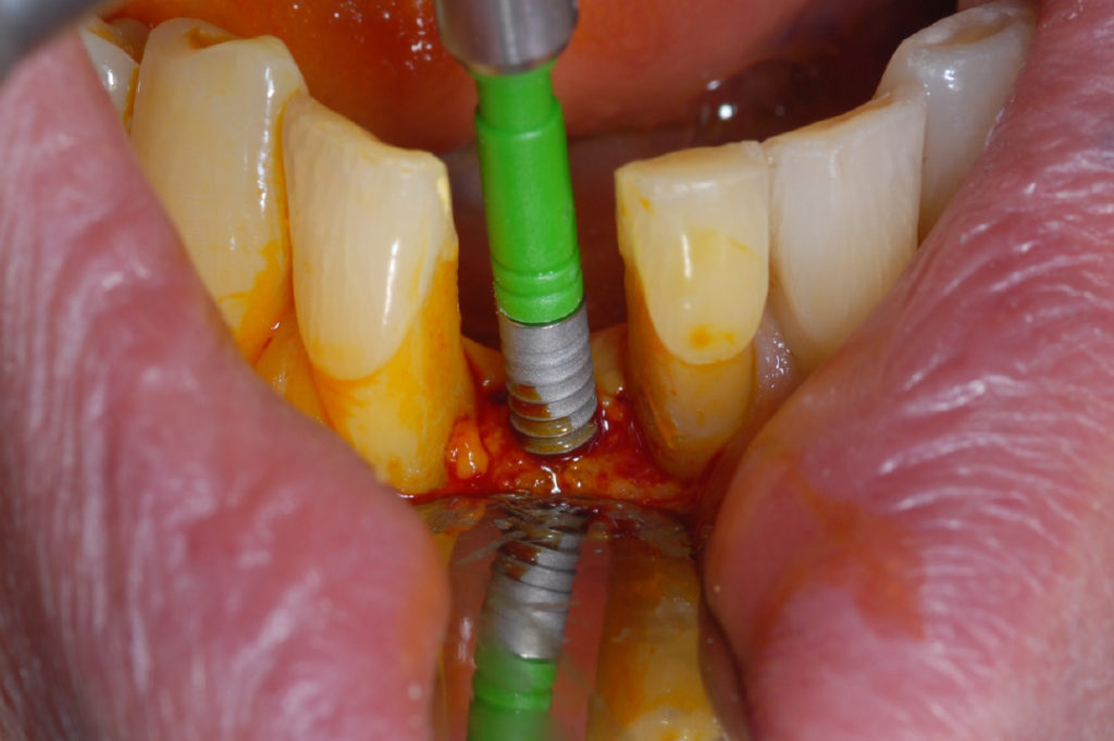
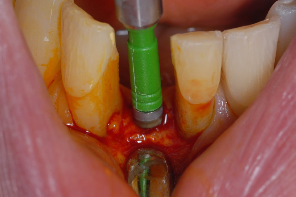
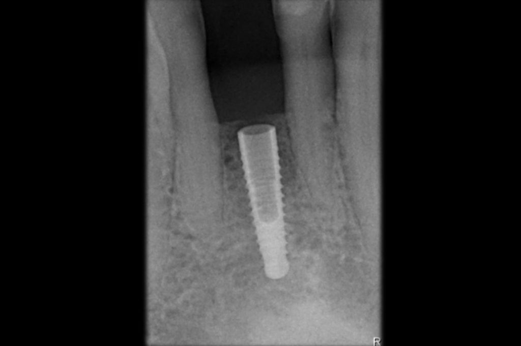
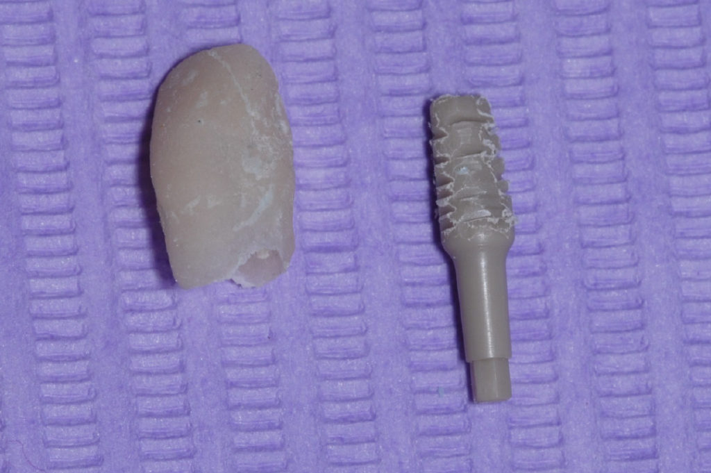
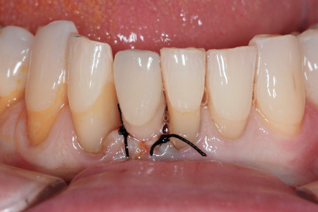
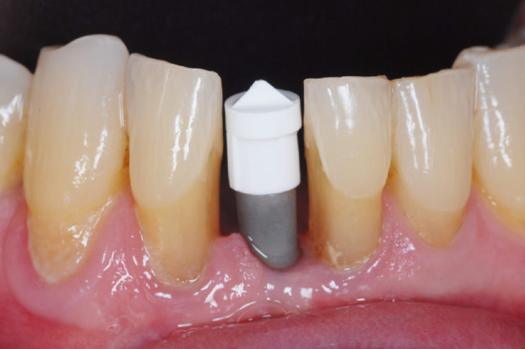
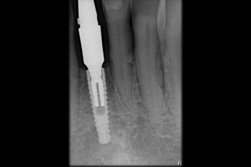
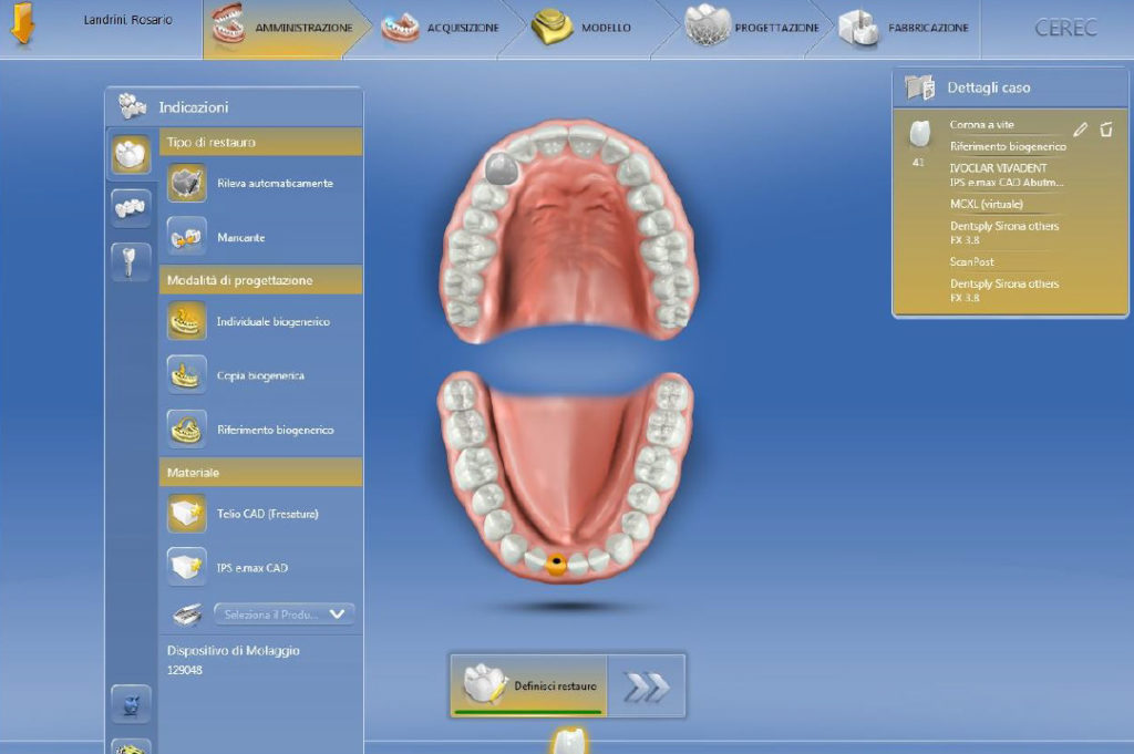
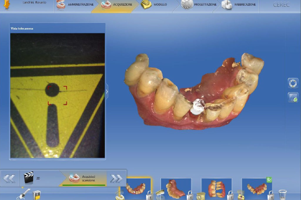
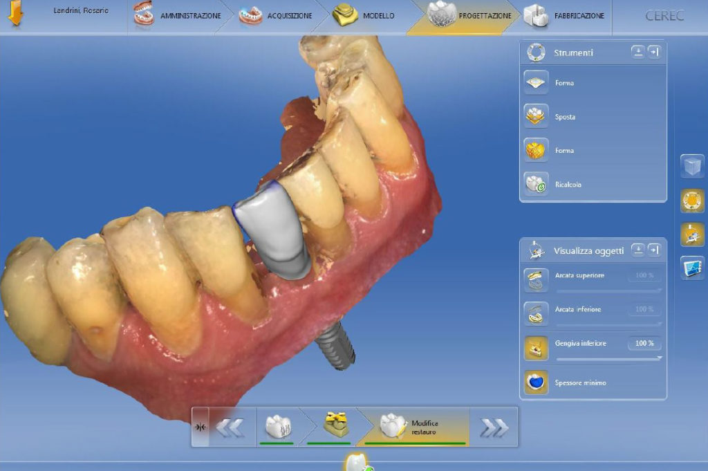
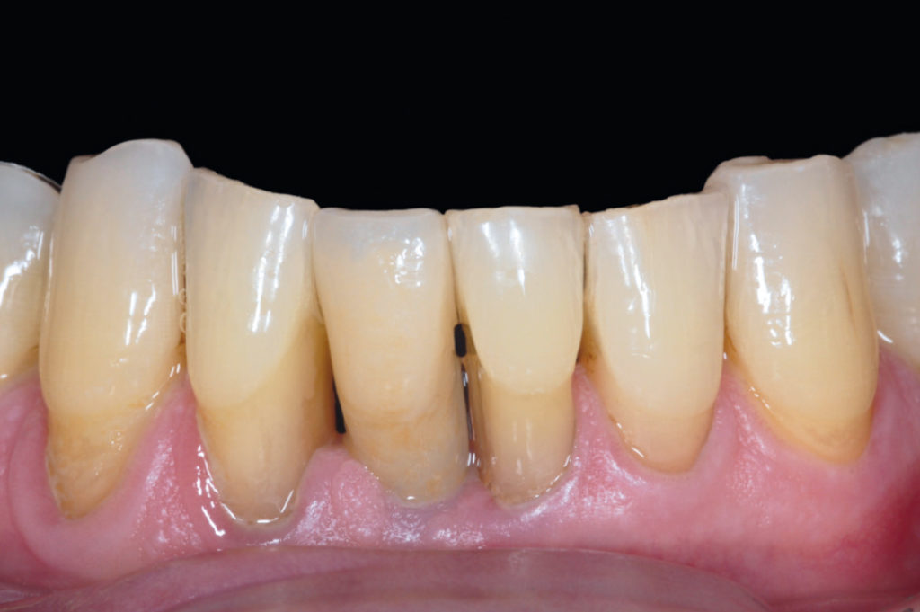
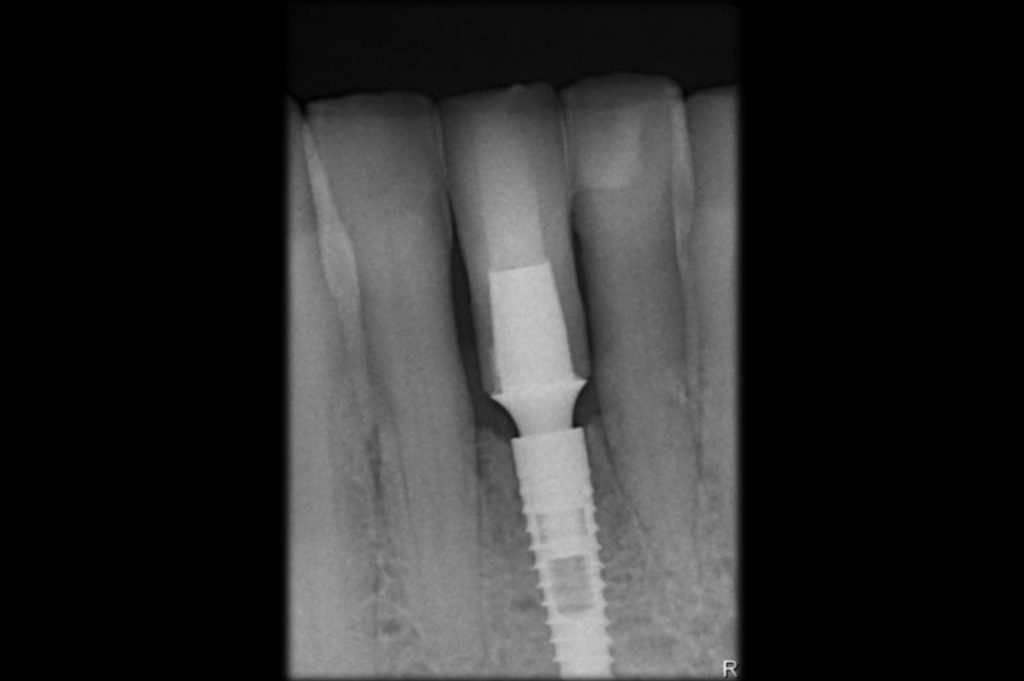
This case shows the insertion and the immediate temporization of a 2.9 x 12 mm XCN implant for the replacement of a mandibular central incisor of a 60-year-old patient. After two and a half months of healing a digital impression was taken and a crown made of lithium disilicate was fabricated and extraorally cemented on a Leone Ti-Base abutment.

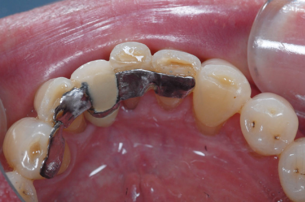
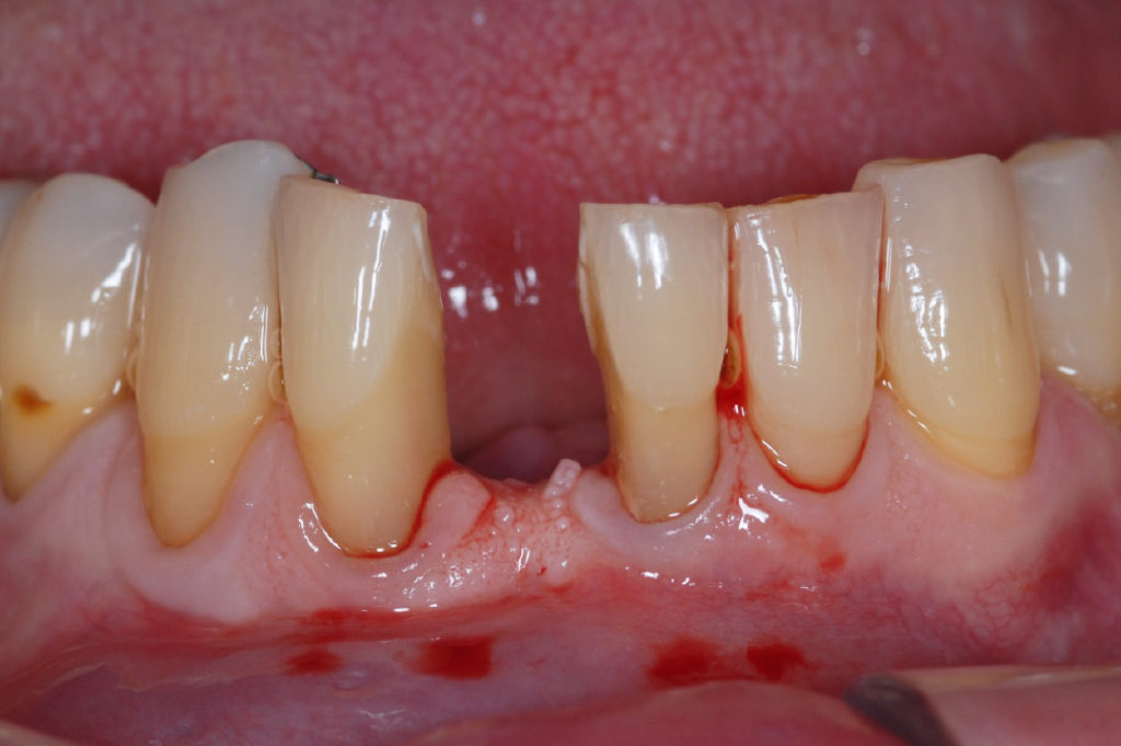
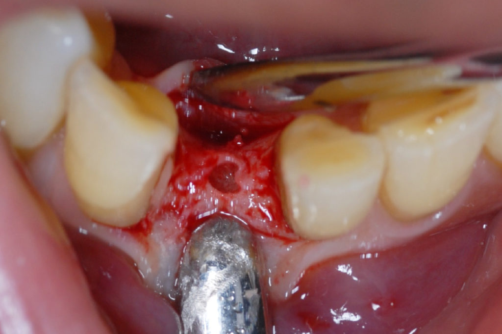
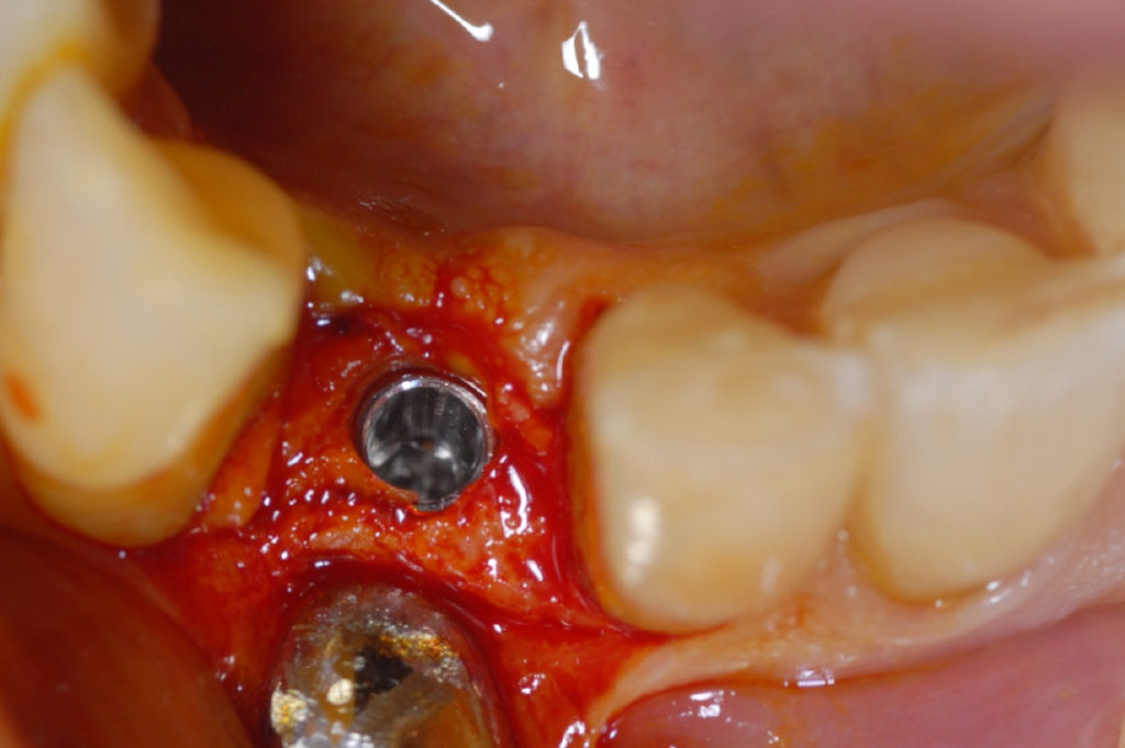
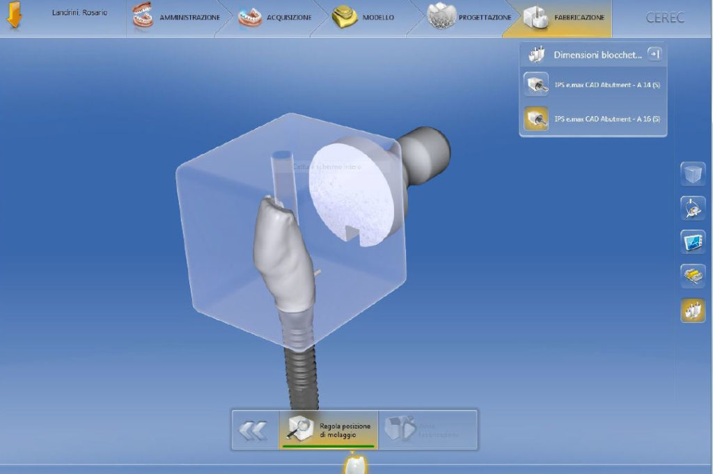
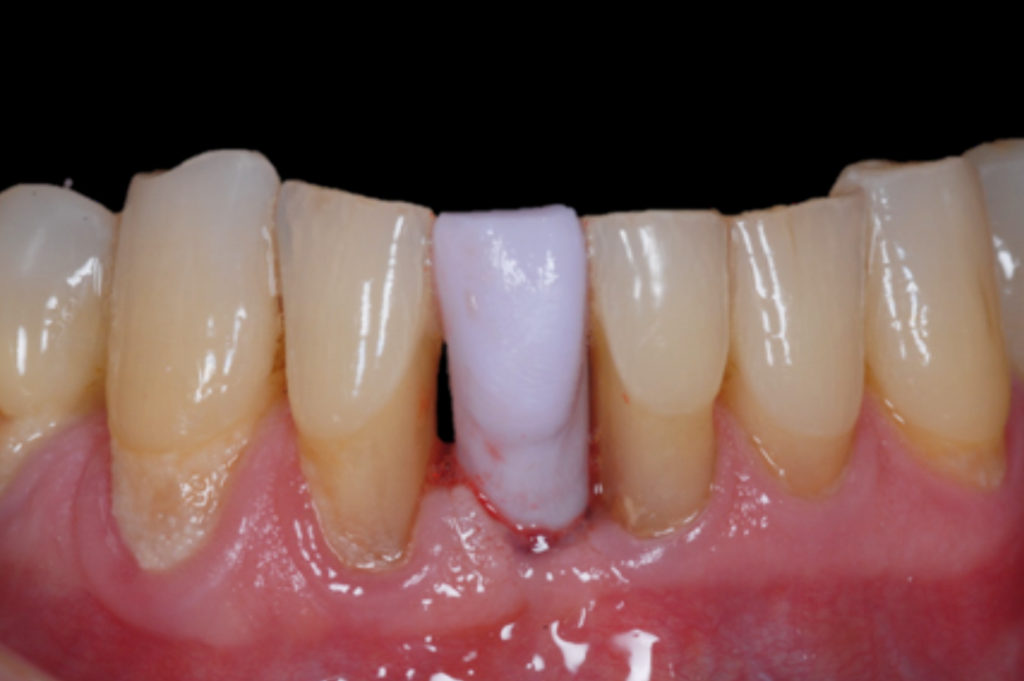
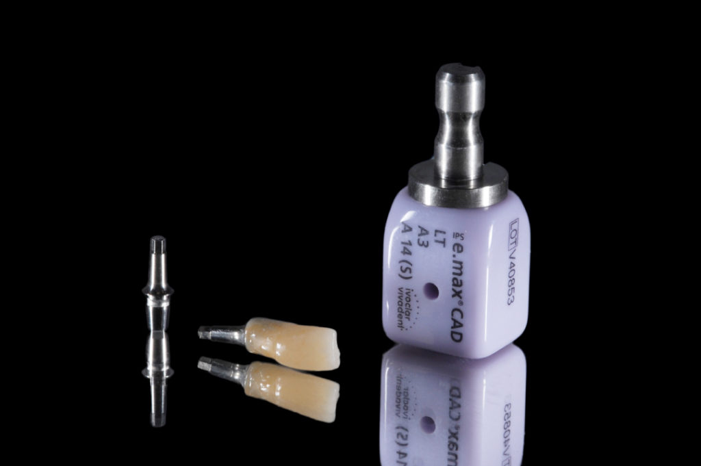
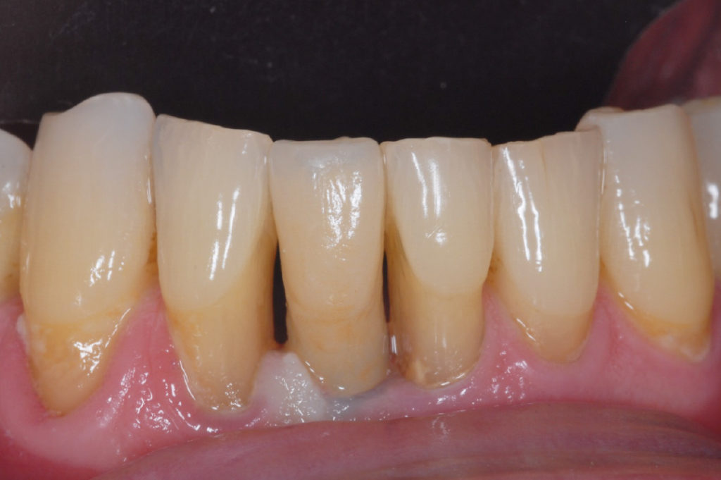
Tooth #41 replaced by Maryland bridge Lingual view of Maryland bridge Preoperative site Access hole to the bone ridge created with lance drill Drilling with 2.2 mm pilot drill for the length of 12 mm Insertion of 2.9 x 12 mm XCN implant Insertion of 2.9 x 12 mm XCN implant Implant in place X-ray to confirm proper seating of implant Prepared temporary abutment and provisional crown for immediate temporization Facial view of immediate temporary restoration Two and a half months later, positioning of scan post coupled with scan body X-ray to confirm proper seating of scan post Starting the scanning process with CEREC, Dentsply Sirona Digital view of scan body recording Planning of crown for tooth #41 Preparation of CAD-CAM milling procedure of the lithium disilicate crown Try-in of lithium disilicate crown and Ti-Base abutment View of Leone Ti-Base abutment, crystallized lithium disilicate crown luted to a Leone Ti-Base abutment and Ivoclar Vivadent e.max CAD block Delivery of the final prosthesis Facial view of final prosthesis after four weeks: proper space around the implant allows for an aesthetically pleasing result X-ray after four weeks confirming optimal implant positioning in relation to adjacent teeth